Інформація призначена тільки для фахівців сфери охорони здоров'я, осіб,
які мають вищу або середню спеціальну медичну освіту.

Підтвердіть, що Ви є фахівцем у сфері охорони здоров'я.

Международный эндокринологический журнал Том 19, №4, 2023

Вернуться к номеру

Поліморфізм TaqI гена рецептора вітаміну D у дітей із соматотропною недостатністю

Авторы: O.V. Bolshova (1), M.O. Ryznychuk (2), D.A. Kvachenyuk (1)
(1) — State Institution “V.P. Komisarenko Institute of Endocrinology and Metabolism of the National Academy of Medical Sciences of Ukraine”, Kyiv, Ukraine
(2) — Bukovinian State Medical University, Chernivtsi, Ukraine

Рубрики: Эндокринология

Разделы: Клинические исследования

Версия для печати


Резюме

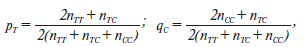
Актуальність. Ядерний рецептор вітаміну D опосередковує більшість біологічних функцій цього вітаміну. Він належить до сімейства рецепторів стероїдних гормонів, ген якого (VDR) розташований на хромосомі 12q13.1. Повідомлялося про генетичну мінливість гена VDR, у якому ідентифіковано понад 470 однонуклеотидних поліморфізмів. Одним з найпоширеніших поліморфізмів у гені VDR є rs731236 (TaqI). Мета дослідження: вивчити поліморфізм TaqI гена VDR у дітей із недостатністю гормону росту (НГР). Матеріали та методи. Визначення TaqI поліморфізму гена VDR (rs731236) проводили за допомогою методу полімеразної ланцюгової реакції з наступним аналізом довжини рестрикційних фрагментів при виявленні їх шляхом електрофорезу в агарозному гелі у 28 дітей препубертатного віку із НГР. ­Результати. У групі хворих із НГР частка гетерозигот Т/С TaqI поліморфізму гена VDR (rs731236) в 1,28 раза вища, ніж серед здорових осіб. Пацієнтів — носіїв генотипу Т/Т та С/С у 0,68 та 0,90 раза менше, ніж у контрольній групі. Наявність гомозиготного генотипу ТТ підвищує ризик розвитку НГР, але невірогідно (відношення шансів (ВШ) = 1,89; 95% довірчий інтервал (ДІ) 0,66–5,39; р = 0,23), а наявність гомозиготного генотипу СС є протективною (ВШ = 0,75; 95% ДІ 0,17–3,22; р = 0,70). При аналізі алелей у пацієнтів із НГР отримані наступні дані: носійство алеля Т поліморфного локусу rs731236 TaqI гена рецептора вітаміну D асоціюється з ризиком соматотропної недостатності (ВШ = 1,24; 95% ДІ 0,65–2,36; р = 0,52), але не досягає статистичної вірогідності. Співвідношення частот алелів (pТ = 0,554, qС = 0,446) практично не відрізняється від 1 : 1, що свідчить про збереження частоти алелів в українській популяції. Висновки. У дітей із НГР частка генотипу Т/С в 1,28 раза вища, ніж у здорових осіб. Наявність гомозиготного генотипу ТТ підвищує ризик розвитку НГР, але невірогідно (ВШ = 1,89; 95% ДІ 0,66–5,39; р = 0,23). Носійство алеля Т поліморфного локусу rs731236 TaqI гена VDR асоціюється з ризиком соматотропної недостатності (ВШ = 1,24; 95% ДІ 0,65–2,36; р = 0,52).

Background. The nuclear receptor for vitamin D mediates most of the biological functions of this vitamin. It belongs to the steroid hormone receptor family, the gene for which (vitamin D receptor — VDR) is located on chromosome 12q13.1. Genetic variability has been reported in the VDR gene, in which more than 470 single nucleotide polymorphisms have been identified. One of the most common polymorphisms in the VDR gene is rs731236 (TaqI). The purpose was to study the TaqI polymorphism of the VDR gene in children with growth hormone deficiency (GHD). Materials and methods. The TaqI polymorphism of the VDR gene (rs731236) was determined using the polymerase chain reaction, followed by analysis of the length of restriction fragments detected by agarose gel electrophoresis in 28 prepubescent children with GHD. Results. In the group of patients with GHD, the proportion of heterozygotes for T/C TaqI polymorphism of the VDR gene (rs731236) is 1.28 times higher than among healthy individuals. There were 0.68 and 0.90 times fewer patients carrying T/T and C/C genotypes than in the control group. The presence of a homozygous TT genotype increases the risk of developing GHD, but not significantly (odds ratio (OR) = 1.89, 95% confidence interval (CI) 0.66–5.39; p = 0.23), and the presence of a homozygous CC genotype is protective (OR = 0.75, 95% CI 0.17–3.22; p = 0.70). When analyzing alleles in patients with GHD, the following data were obtained: carriage of the T allele for the polymorphic loci TaqI rs731236 of the VDR gene is associated with the risk of GHD (OR = 1.24, 95% CI 0.65–2.36; p = 0.52) but not significantly. The ratio of allele (pТ = 0.554, qС = 0.446) frequencies practically does not differ from 1 : 1, which indicates the preservation of allele frequencies in the Ukrainian population. Conclusions. In children with GHD, the proportion of the T/C genotype is 1.28 times higher than in the group of healthy persons. The presence of a homozygous TT genotype increases the risk of developing GHD but not significantly (OR = 1.89, 95% CI 0.66–5.39; p = 0.23). Carriage of the T allele for the polymorphic locus TaqI rs731236 of the VDR gene is associated with the risk of the growth hormone deficiency (OR = 1.24, 95% CI 0.65–2.36; p = 0.52) but not significantly.


Ключевые слова

соматотропна недостатність; діти; TaqI поліморфізм гена рецептора вітаміну D; розподіл генотипів

growth hormone deficiency; children; TaqI polymorphism of the vitamin D receptor gene; distribution of genotypes

Introduction

Vitamin D, which is a powerful regulator of bone and calcium homeostasis, as well as immunomodulation, cell differentiation and replication in various target tissues, has attracted great interest from the scientists all over the world in recent years. Vitamin D receptor (VDR) gene polymorphisms are associated with multiple features, and phenotypes of disease such as primary hyperparathyroidism, Graves’ disease, type 1 diabetes mellitus, and osteoporosis [1].
The nuclear vitamin D receptor mediates most of the biological functions of this vitamin [2]. It belongs to the steroid hormone receptor family whose gene (VDR) is located on chromosome 12q13.1 [3]. VDR has eight exons and six introns, located in genetically active regions, with promoter segments [2].
Genetic variability has been reported in the VDR gene, in which more than 470 single nucleotide polymorphisms have been identified. The two most common polymorphisms in the VDR gene are rs2228570 (FokI) and rs731236 (TaqI) [4]. These polymorphisms in the VDR gene have been described using FokI and TaqI restrictases. The TaqI polymorphism (rs731236) is located in exon 9, T/t (cytosine → thymine) as a silent mutation [5], whereas FokI is located in exon 2 and causes the replacement of T nucleotides with C [6].
It has also been shown that the bone mass control, as well as bone diseases, are associated with a genetic polymorphism of the VDR gene [6, 7].
The TaqI polymorphism is a T/C nucleotide substitution (ATT to ATC) resulting in a synonymous substitution of codon 352 (isoleucine) in exon IX [8].
The polymorphism frequencies depend on ethnicity, so the comparison of these frequencies using a chi-square test with other populations is statistically significant for FokI and TaqI [9].
Disagreements between studies on genetic risks that may be associated with the genetic heterogeneity of the VDR gene, exogenous population influence and complex gene interactions exist to this day [10]. Therefore, the study of VDR gene polymorphisms in certain population groups is quite actual.
The purpose of the study was to study the TaqI polymorphism of the VDR gene in children with SD.

Materials and methods

A genetic study was carried out in 28 children with GHD who were treated at the State Institution “V.P. Komisarenko Institute of Endocrinology and Metabolism of the National Academy of Medical Sciences of Ukraine”.
The following indexes were taken into account: the patient’s gender and age, anthropometric data, serum vitamin D level (the summer months of patient recruitment were excluded), bone age, the level of growth hormone (GH) after stimulation tests (clonidine, insulin), the levels of insulin-like growth factor-1 (IGF-1), serum level of total and ionized calcium.
The average age of the children (21 boys, 7 girls) inclu–ded in the study was 10.86 ± 3.15 years. The average growth retardation was –2.34 ± 0.85 SDS. By the time of the exami–nation, all patients had euthyroidism. The study included children who had not received calcium and vitamin D supplements for ≥ 6 months. Children with GHD had a significant decrease in the level of IGF-1 (from 22.83 to 93.04 ng/ml). There were taken 57 children for the control group.
To determine polymorphic variants of TaqI (rs731236), modified protocols of the VDR gene, that predict involving the use of oligonucleotide primers in the polymerase chain reaction (PCR) technique and subsequent restriction fragment length polymorphism (RFLP) when they were detected by agarose gel electrophoresis were used. For genotyping, venous blood was collected under sterile conditions in 2.7 ml monovette with potassium salt of ethylenediaminetetraacetic acid (Sarstedt, Germany), which served as an anticoagulant. Initially, DNA was eliminated from peripheral blood using the commercial Quick-DNA Miniprep Plus Kit (Zymo Research, USA). The target gene region was amplified using the corresponding specific primer (Metabion, Germany).
Amplification was performed using a commercial kit called DreamTaq Green PCR Master Mix (2X) (Thermo Scientific, USA) and specific primers (Metabion, Germany). A FlexCycler amplifier (Analytik Jena, Germany) was used for the corresponding thermal PCR regime.
The amplified product (amplicons) was subjected to hydrolysis with TaqI restriction endonucleases (10 U/μl) (Thermo Scientific, USA). Restriction hydrolysis was performed according to the manufacturer’s instructions in a dry block heater for 16 hours at 65 °C to study the TaqI VDR gene polymorphism.
The VDR TaqI polymorphic variant did not require thermal inactivation according to the manufacturer’s instructions. The state of the restriction fragments of the VDR gene (TaqI) was analyzed in a 2% agarose gel (CSL-AG500 agarose, Cleaver Scientific Ltd, Great Britain) using the ethi–dium bromide as a dye. To assess the size of the fragments, a molecular weight marker was introduced — GeneRuler 100 bp DNA Ladder (Thermo Scientific, USA) and then visua–lized in a transilluminator stained with ethidium bromide using the Vitran computer program.
The enhancers for TaqI T/C polymorphism the VDR gene (rs731236) were subjected to hydrolytic cleavage in the presence of the 5’-GAATGCN↓-3’ restriction site, resulting in the formation of restikts with a molecular weight of 496 and 249 bp — TT genotype.
The restriction site disappeared upon nucleotide substitution from T to C, if the size of the amplified DNA fragments after interaction with the restriction nuclease had a molecular weight of 295, 249 and 201 bp, then the CC genotype was recorded. Accordingly, in the heterozygous genotype (TC) all four types of fragments were observed simultaneously: 496, 295, 249 and 201 bp.
Statistical processing of the research results was carried out using Microsoft Excel statistical programs.
The distribution of genotypes in the groups of patients and healthy people was compared according to the Hardy-Weinberg principle (χ2):
р2 + 2pq + q2 = 100 %,
where p2 is the frequency with which carriers of the TT geno–type occur, 2pq are of the TC genotypes, and q2 are of the CC genotype.
Frequencies of pТ and qС alleles were calculated as:
where n is the number of persons with a certain genotype.
The odds ratio (OR) was calculated:
where a is the presence of GHD of the studied sign, b is the presence of GHD and the absence of the studied sign, c is healthy persons and the absence of the studied sign, d is healthy persons and the presence of the studied sign.
A confidence interval (CI) was calculated at the 95% significance level for the OR. If the odds ratio was less than 1, then the risk decreased, if is was equal to 1, then there was no risk, if more than 1, then the risk was present [11]. 
All data were analyzed by nonparametric methods of variational statistics using the computer program MedCalc (2006).
The study was conducted in accordance with the basic principles of bioethics of the Council of Europe Convention on Human Rights and Biomedicine (April 4, 1997), the Helsinki Declaration of the World Health Association on the ethical principles of medical research involving human subjects (1964–2013). The Biomedical Ethics Commission of the State Institution “V.P. Komisarenko Institute of Endocrinology and Metabolism of the National Academy of Medical Sciences of Ukraine” did not reveal any violations of moral and legal norms during the study. Informed consent was obtained from the participants and their parents.

Results

In GHD patients the proportion of the T/C genotype is 1.28 times higher than in the group of healthy persons. There are 0.68 and 0.90 times fewer patients-carriers the T/T and C/C genotypes than in the control group (Table 1). The presence of a homozygous TT genotype increases the risk of developing GHD, but not significantly (OR = 1.89, 95% CI 0.66–5.39; p = 0.23), and the presence of a homozygous CC genotype is protective (OR = 0.75, 95% CI 0.17–3.22; p = 0.70).
When analyzing alleles in patients with GHD, the following data were obtained: the carrier of the T allele of the polymorphic locus TaqI (rs731236) of the VDR gene is associated with the risk of GH deficiency (OR = 1.24, 95% CI 0.65–2.36; p = 0.52) but not significantly.
The main allele in the control group is T (pТ = 0.605), as well as in the group of GHD patients (pТ = 0.554). The frequency of the minor C allele in GHD patients (qС = 0.395) almost does not differ from the group of healthy individuals (qС = 0.446; Table 2).
The ratio of allele frequencies (pТ = 0.554, qС = 0.446) practically does not differ from the 1 : 1 ratio, which indicates the preservation of the allele frequencies in the Ukrainian population.
The allele frequencies in GHD patients differed significantly from those in the control group, but the distribution of genotypes corresponded to the Hardy-Weinberg equilibrium (Table 3).
In the cohort of Ukrainian children, as well as in the control group, heterozygous T/С carriers prevailed.

Discussion

Over the past decades, the frequency as well as the budgets for vitamin D testing has increased significantly in many countries [13]. Prescribing vitamin D to patients is the result of many promising clinical studies linking low levels of vitamin D to the risk of developing certain diseases, namely cardiovascular pathology, growth pathology, cancer, diabetes mellitus [14]. The number of published papers on vitamin D, as well as diseases associated with its deficiency, is increasing daily [15].
Transportation of vit D and its metabolites occurs in a bound form with proteins: 85 % with vitamin D binding protein and 15 % with albumin [16]. When this complex reaches the target cells, vitamin D is released from proteins and enters the cell to interact with the nuclear VDR. The latter can be found in various tissues and cells. VDR functions as a transcription factor. The liganded VDR binds to the retinoid X receptor to form a heterodimeric complex that can activate or repress gene expression by binding to elements in the VDR promoter region of the regulated gene (vitamin D response element), initiating the formation of nuclear transcription factors [15].
The TaqI polymorphism showed a strong correlation with vitamin D levels (P = 0.021) when analyzed by the chi-square test. The TaqI SNP is reported to be in linkage disequilibrium with BsmI and ApaI and may be considered as a marker of bone mineral density in humans [6].
The association of the TaqI genotype with vitamin D levels may be explained by the effect of polymorphisms in the VDR gene on calcium metabolism, which in turn plays an important role in the feedback mechanism of vitamin D levels. It has been shown that the TaqI polymorphism can be in non-equilibrium linkage with another marker that significantly affects the level of vitamin D [17]. In addition, individual homozygotes with the C allele of the TaqI polymorphism showed a higher risk of osteoporosis in the populations of Saudi Arabia and the Caucasus [18, 19].
Functional studies of each polymorphism in the VDR gene are ongoing, and an association between VDR polymorphisms and height has been found in several other studies, requiring further investigation [20, 21].

Conclusions

In GHD children, the proportion of the T/C genotype is 1.28 times higher than in the group of healthy persons. The presence of a homozygous TT genotype increases the risk of developing GHD, but not significantly (OR = 1.89, 95% CI 0.66–5.39; p = 0.23).
The carriage of the T allele of the polymorphic locus TaqI (rs731236) of the VDR gene is associated with the risk of GH deficiency (OR = 1.24, 95% CI 0.65–2.36; p = 0.52) but not significantly.
 
Received 10.02.2023
Revised 19.04.2023
Accepted 04.05.2023

Список литературы

  1. Wang Y., Zhu J., DeLuca H.F. Where is the vitamin D receptor? Arch. Biochem. Biophys. 2012 Jul 1. 523(1). 123-33. doi: 10.1016/j.abb.2012.04.001.
  2. Tuoresmäki P., Väisänen S., Neme A., Heikkinen S., Carlberg C. Patterns of genome-wide VDR locations. PLoS One. 2014 Apr 30. 9(4). e96105. doi: 10.1371/journal.pone.0096105.
  3. Uitterlinden A.G., Fang Y., van Meurs J.B., van Leeu–wen H., Pols H.A. Vitamin D receptor gene polymorphisms in relation to vitamin D-related disease states. J. Steroid Biochem. Mol. Biol. 2004 May. 89–90 (1–5). 187-93. doi: 10.1016/j.jsbmb.2004.03.083.
  4. Pankiv V.I., Yuzvenko T.Yu., Pankiv I.V. Type 2 diabetes mellitus and subclinical hypothyroidism: focusing on the role of cholecalciferol. Problems of Endocrine Pathology. 2019. 2. 46-51. doi: 10.21856/j-PEP.2019.2.07.
  5. Cieślińska A., Kostyra E., Chwała B., Moszyńska-Dumara M., Fiedorowicz E. et al. Vitamin D receptor gene polymorphisms associa–ted with childhood autism. Brain Sci. 2017 Sep 9. 7(9). 115. doi: 10.3390/brainsci7090115.
  6. Bhanushali A.A., Lajpal N., Kulkarni S.S., Chavan S.S., Bagadi S.S., Das B.R. Frequency of FOKI and TAQI polymorphism of vitamin D receptor gene in Indian population and its association with 25-hydroxyvitamin D levels. Indian J. Hum. Genet. 2009 Sep. 15(3). 108-13. doi: 10.4103/0971-6866.60186.
  7. Osman E., Al Anouti F., El Ghazali G., Haq A., Mirgani R., Al Safar H. Frequency of rs731236 (Taql), rs2228570 (Fok1) of vitamin-D receptor (VDR) gene in Emirati healthy population. Meta Gene. 2015 Sep 15. 6. 49-52. doi: 10.1016/j.mgene.2015.09.001.
  8. Hustmyer F.G., DeLuca H.F., Peacock M. ApaI, BsmI, EcoRV and TaqI polymorphisms at the human vitamin D receptor gene locus in Caucasians, blacks and Asians. Hum. Mol. Genet. 1993 Apr. 2(4). 487. doi: 10.1093/hmg/2.4.487.
  9. Emmanouilidou E., Galli-Tsinopoulou A., Kyrgios I., Gbandi E., Goulas A. Common VDR polymorphisms and idiopathic short stature in children from northern Greece. Hippokratia. 2015 Jan-Mar. 19(1). 25-9.
  10. Steppe L., Bülow J., Tuckermann J., Ignatius A., Haffner-Luntzer M. Bone mass and osteoblast activity are sex-dependent in mice lacking the estrogen receptor α in chondrocytes and osteoblast progenitor cells. Int. J. Mol. Sci. 2022 Mar 7. 23(5). 2902. doi: 10.3390/ijms23052902.
  11. Fletcher D., Faddy M. Confidence Intervals for Expec–ted Abundance of Rare Species. Journal of Agricultural, Biological, and Environmental Statistics. 2007. 12(3). 315-324. Available from: http://www.jstor.org/stable/27595645.
  12. Stranger B.E., Stahl E.A., Raj T. Progress and promise of genome-wide association studies for human complex trait genetics. Genetics. 2011 Feb. 187(2). 367-83. doi: 10.1534/genetics.110.120907.
  13. Nikolac Gabaj N., Unic A., Miler M., Pavicic T., Culej J., Bolanca I. et al. In sickness and in health: pivotal role of vitamin D. Biochem. Med. (Zagreb). 2020 Jun 15. 30(2). 020501. doi: 10.11613/BM.2020.020501.
  14. Zhao S., Gardner K., Taylor W., Marks E., Goodson N. Vitamin D assessment in primary care: changing patterns of tes–ting. London J. Prim. Care (Abingdon). 2015. 7(2). 15-22. doi: 10.1080/17571472.2015.11493430.
  15. Yang A., Lv Q., Chen F., Wang D., Liu Y., Shi W. Identification of recent trends in research on vitamin D: a quantitative and co-word analysis. Med. Sci. Monit. 2019 Jan 22. 25. 643-55. doi: 10.12659/MSM.913026.
  16. Haussler M.R., Whitfield G.K., Kaneko I., Haussler C.A., Hsieh D. et al. Molecular mechanisms of vitamin D action. Calcif. Tissue Int. 2013 Feb. 92(2). 77-98. doi: 10.1007/s00223-012-9619-0.
  17. Singh I., Lavania M., Pathak V.K., Ahuja M., Turan–kar R.P. et al. VDR polymorphism, gene expression and vitamin D levels in leprosy patients from North Indian population. PLoS Negl. Trop. Dis. 2018 Nov 27. 12(11). e0006823. doi: 10.1371/journal.pntd.0006823.
  18. Kow M., Akam E., Singh P., Singh M., Cox N. et al. Vitamin D receptor (VDR) gene polymorphism and osteoporosis risk in White British men. Ann. Hum. Biol. 2019 Aug. 46(5). 430-3. doi: 10.1080/03014460.2019.1659851.
  19. Jiang L.L., Zhang C., Zhang Y., Ma F., Guan Y. Associations between polymorphisms in VDR gene and the risk of osteoporosis: a meta-analysis. Arch. Physiol. Biochem. 2022 Dec. 128(6). 1637-44. doi: 10.1080/13813455.2020.1787457.
  20. Haussler M.R., Whitfield G.K., Haussler C.A., Hsieh J.C., Thompson P.D. et al. The nuclear vitamin D receptor: biological and molecular regulatory properties revealed. J Bone Miner. Res. 1998. 13(3). 325-49. doi: 10.1359/jbmr.1998.13.3.325.
  21. Xiong D.H., Xu F.H., Liu P.Y., Shen H., Long J.R. еt al. Vitamin D receptor gene polymorphisms are linked to and associated with adult height. J. Med. Genet. 2005. 42(3). 228-34. doi: 10.1136/jmg.2004.024083.

Вернуться к номеру