
Международный эндокринологический журнал Том 22, №1, 2026
Вернуться к номеру
Гістологічні орієнтири для вибору терапевтичного маршруту в пацієнток із перехресним синдромом генітального ендометріозу та доброякісного захворювання молочної залози
Авторы: S.V. Konovalenko (1), A.D. Neborets (2), V.V. Protsenko (3), D.S. Androsov (4), A.V. Khmel (5), A.S. Lunko (6)
(1) - R.E. Kavetsky Institute of Experimental Pathology, Oncology and Radiobiology of NASU, Kyiv, Ukraine
(2) - National Cancer Institute, Kyiv, Ukraine
(3) - State Institution “Institute of Traumatology and Orthopedics of NAMS of Ukraine”, Kyiv, Ukraine
(4) - O.F. Herbachevsky Zhytomyr Regional Clinical Hospital, Zhytomyr, Ukraine
(5) - Specialized Mammological Center, Kyiv, Ukraine
(6) - Odesa National Medical University, Odesa, Ukraine
Рубрики: Эндокринология
Разделы: Клинические исследования
Версия для печати
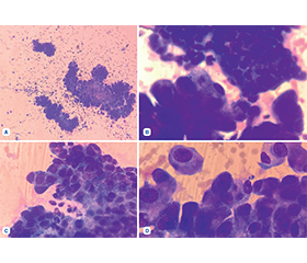
Актуальність. Генітальний ендометріоз (ГЕ) залишається дуже поширеним захворюванням, яке зустрічається в 0,5–5 % фертильних та 25–40 % безплідних жінок. Доброякісні захворювання молочної залози (ДЗМЗ) поширені протягом усього життя, від раннього репродуктивного віку до постменопаузального періоду, що робить їх потенційною проблемою для здоров’я великої кількості жінок. Гістологічні характеристики тканин ендометрію та молочної залози в пацієнток, які страждають на перехресний синдром ГЕ та ДЗМЗ, становлять інтерес, оскільки ця патологія несе підвищений ризик раку ендометрія, яєчників і молочної залози. Мета: вивчити та виявити найбільш характерні особливості ендометріоїдної тканини й тканини гіперплазії проток молочної залози в жінок із перехресним синдромом ГЕ та ДЗМЗ за допомогою світлової мікроскопії. Матеріали та методи. Досліджено гістологічні препарати ендометріоїдних вогнищ і тканин гіперплазії молочної залози 148 жінок із перехресним синдромом ГЕ та ДЗМЗ. Матеріал був отриманий методом тонкоголкової аспіраційної пункційної біопсії, яку проводили під ультразвуковим контролем. Цитологічний аналіз та мікрофотографування виконували за допомогою мікроскопа Olympus CX23 із різним збільшенням (×40, ×100 (занурення)). Результати. Дослідження показало, що у відібраній когорті пацієнток із перехресним синдромом ГЕ та ДЗМЗ переважна більшість гістологічних зразків демонстрували проліферативну активність мезотеліальних клітин. Водночас морфологічні ознаки атипії клітинних елементів атипової гіперплазії проток спостерігалися в 47,3 % випадків. Клітини з ознаками злоякісного переродження не були виявлені в жодному з наданих зразків тканини молочної залози. Важливо зазначити, що в 15 % пацієнток у зразках ендометрія діагностовано гістологічні ознаки злоякісного новоутворення, що призвело до їхнього негайного направлення до гінеколога-онколога для хірургічного втручання та необхідної ад’ювантної терапії. Висновки. У прогнозі клінічного перебігу перехресного синдрому ГЕ та ДЗМЗ ключову роль відіграють гістологічне підтвердження діагнозу та ознаки атипії у зразках біопсії. Виявлення атипових клітин вимагає клінічної консультації за участю гінеколога, мамолога та гінеколога-онколога, під час якої приймається рішення щодо хірургічного втручання та необхідної фармакотерапії.
Background. Genital endometriosis (GE) remains a very common disease, occurring in 0.5–5 % of fertile women and in 25–40 % of infertile women. Benign breast diseases (BBD) are common throughout life, from early reproductive age to the postmenopausal period, making them a potential health problem for a large number of women. The histological characteristics of endometrial and breast tissues in patients suffering from the overlap syndrome of GE and BBD are of interest, since this condition carries an increased risk of endometrial, ovarian and breast cancer. The purpose was to study and identify the most characteristic features of endometrioid tissue and ductal hyperplasia tissue of the breast in women with overlap syndrome of GE and BBD using light microscopy. Materials and methods. Histological preparations of endometrioid foci and breast hyperplasia tissues of 148 women with overlap syndrome of GE and BBD were studied. The material was obtained by the method of fine-needle aspiration puncture biopsy, which was performed under ultrasound control. Cytological analysis and microphotography were performed using an Olympus CX23 microscope with different magnifications (×40, ×100 (immersion)). Results. The study showed that in the selected cohort of patients with overlap syndrome of GE and BBD, most histological samples showed proliferative activity of mesothelial cells. At the same time, morphological signs of atypia of cellular elements of atypical ductal hyperplasia were observed in 47.3 % of histological samples of breast tissue. Cells with signs of malignant transformation were not found in any of the provided breast tissue samples. It is important to note that in 15 % of patients, histological signs of malignancy were found in endometrial samples, which led to their immediate referral to a gynecological oncologist for surgical intervention and the necessary adjuvant therapy. Conclusions. In the prognosis of the clinical course of GE and BBD overlap syndrome, histological confirmation of the diagnosis and detection of signs of atypia in biopsy samples play a key role. The finding of atypical cells requires clinical consultation with the participation of a gynecologist, a mammologist and a gynecological oncologist, during which a decision is made regarding surgical intervention and the necessary pharmacotherapy.
ендометріоз; доброякісне захворювання молочної залози; атипова гіперплазія проток; перехресний синдром; ендометріоїдна карцинома; рак молочної залози; метаболічний синдром; кісткові метастази
endometriosis; benign breast disease; atypical ductal hyperplasia; overlap syndrome; endometrioid carcinoma; breast cancer; metabolic syndrome; bone metastases
Для ознакомления с полным содержанием статьи необходимо оформить подписку на журнал.
- Mariadas H, Chen JH, Chen KH. The Molecular and Cellular Mechanisms of Endometriosis: From Basic Pathophysiology to Clinical Implications. Int J Mol Sci. 2025 Mar 10;26(6):2458. doi: 10.3390/ijms26062458.
- Borojerdi ASD, Welchowski T, Peng WM, Buchen A, Novak N, Haidl G, et al. Human spermatozoa of male patients with subfertility express the interleukin-6 receptor. Andrologia. 2020;52:e13511. doi: 10.1111/and.13511.
- Lin SC, Li WN, Lin SC, Hou HT, Tsai YC, Lin TC, et al. Targeting Yap1 ameliorates progesterone resistance in endometriosis. Hum Reprod. 2023;38:1124-34. doi: 10.1093/humrep/dead071.
- Laganà AS, Salmeri FM, Ban Frangež H, Ghezzi F, Vrtač–nik-Bokal E, Granese R. Evaluation of M1 and M2 macrophages in ovarian endometriomas from women affected by endometriosis at different stages of the disease. Gynecol Endocrinol. 2020;36:441-4. doi: 10.1080/09513590.2019.1683821.
- Di Spiezio Sardo A, Becker CM, Renner SP, Suvitie PA, Tarriel JE, Vannuccini S, et al. Management of women with endometriosis in the 21st century. Curr Opin Obstet Gynecol. 2025 Jun 1;37(3):149-157. doi: 10.1097/GCO.0000000000001027.
- Ball E, Khan KS. Recent advances in understanding and mana–ging chronic pelvic pain in women with special consideration to endometriosis. F1000Res. 2020;9:F1000, Faculty Rev-83. doi: 10.12688/f1000research.20750.1.
- Parasar P, Ozcan P, Terry KL. Endometriosis: Epidemiology, Diagnosis and Clinical Management. Curr Obstet Gynecol Rep. 2017;6(1):34-41. doi: 10.1007/s13669-017-0187-1.
- Wang Y, Nicholes K, Shih IM. The Origin and Pathogenesis of Endometriosis. Annu Rev Pathol. 2020;15:71-95. doi: 10.1146/annurev-pathmechdis-012419-032654.
- Rolla E. Endometriosis: advances and controversies in classification, pathogenesis, diagnosis, and treatment. F1000Res. 2019;8:F1000, Faculty Rev-529. doi: 10.12688/f1000research.14817.1.
- Sieberg CB, Lunde CE, Borsook D. Endometriosis and pain in the adolescent — striking early to limit suffering: A narrative review. Neurosci Biobehav Rev. 2020;108:866-876. doi: 10.1016/j.neubio–rev.2019.12.004.
- Ruszała M, Dłuski DF, Winkler I, Kotarski J, Rechber–ger T, Gogacz M. The State of Health and the Quality of Life in Women Suffering from Endometriosis. Journal of Clinical Medicine. 2022;11(7):2059. doi: 10.3390/jcm11072059.
- Stachs A, Stubert J, Reimer T, Hartmann S. Benign Breast Disease in Women. Dtsch Arztebl Int. 2019 Aug 9;116(33–34):565-574. doi: 10.3238/arztebl.2019.0565.
- Johansson A, Christakou AE, Iftimi A, Eriksson M, Tapia J, Skoog L, et al. Characterization of Benign Breast Diseases and Association With Age, Hormonal Factors, and Family History of Breast Cancer Among Women in Sweden. JAMA Netw Open. 2021 Jun 1;4(6):e2114716. doi: 10.1001/jamanetworkopen.2021.14716.
- Burke A, O’Driscoll J, Abubakar M, Bennett KE, Carmody E, Flanagan F, et al. A systematic review of determinants of breast cancer risk among women with benign breast disease. NPJ Breast Cancer. 2025 Feb 15;11(1):16. doi: 10.1038/s41523-024-00703-w.
- Figueroa JD, Gierach GL, Duggan MA, et al. Risk factors for breast cancer development by tumor characteristics among women with benign breast disease. Breast Cancer Res. 2021;23:34. doi: 10.1186/s13058-021-01410-1.
- Rubio IT, Wyld L, et al. European guidelines for the diagnosis, treatment and follow-up of breast lesions with uncertain malignant potential (B3 lesions) developed jointly by EUSOMA, EUSOBI, ESP (BWG) and ESSO. Eur J Surg Oncol. 2024 Jan;50(1):107292. doi: 10.1016/j.ejso.2023.107292.
- Siegel RL, Kratzer TB, Giaquinto AN, Sung H, Jemal A. Cancer statistics, 2025. CA Cancer J Clin. 2025 Jan-Feb;75(1):10-45. doi: 10.3322/caac.21871.
- Brockton NT, Cook LS, Magliocco AM, Shemanko CS, Vogel HJ, еt al. The Breast to Bone (B2B) Cohort Study to Prevent, Detect and Improve Treatment of Metastatic Disease: Baseline Assessment, Description and Progress. International Journal of Environmental Research and Public Health. 2025;22(2):242. doi: 10.3390/ijerph22020242.
- Eby PR, Ochsner JE, DeMartini WB, Allison KH, Peacock S, Lehman CD. Frequency and upgrade rates of atypical ductal hyperplasia diagnosed at stereotactic vacuum-assisted breast biopsy: 9- versus 11-gauge. AJR. 2009;192:229-234. doi: 10.2214/AJR.08.1342.
- Schiaffino S, Calabrese M, Melani EF, Trimboli RM, Cozzi A, Carbonaro LA, Di Leo G, Sardanelli F. Upgrade Rate of Percutaneously Diagnosed Pure Atypical Ductal Hyperplasia: Systematic Review and Meta-Analysis of 6458 Lesions. Radiology. 2020;294:76-86. doi: 10.1148/radiol.2019190748.
- Burbank F. Stereotactic breast biopsy of atypical ductal hyperplasia and ductal carcinoma in situ lesions: Improved accuracy with directional, vacuum-assisted biopsy. Radiology. 1997;202:843-847. doi: 10.1148/radiology.202.3.9051043.
- Gomes DS, Porto SS, Balabram D, Gobbi H. Inter-obser–ver variability between general pathologists and a specialist in breast pathology in the diagnosis of lobular neoplasia, columnar cell lesions, atypical ductal hyperplasia and ductal carcinoma in situ of the breast. Diagn Pathol. 2014;9:121. doi: 10.1186/1746-1596-9-121.
- Tozbikian G, Brogi E, Vallejo CE, Giri D, Murray M, Catalano J, et al. Atypical Ductal Hyperplasia Bordering on Ductal Carcinoma In Situ. Int J Surg Pathol. 2017;25:100-107. doi: 10.1177/1066896916662154.
- Amin A, Winblad O, Zupon A, Fan F, Tawfik O, Wick J, et al. Atypical ductal hyperplasia on percutaneous breast biopsy: Scoring system to identify the lowest risk for upgrade. Res Sq. 2021. doi: 10.21203/rs.3.rs-388478/v1.
- Istrate-Ofiţeru A-M, Mogoantă CA, Zorilă G-L, Roşu G-C, Drăguşin RC, Berbecaru E-I-A, et al. Clinical Characteristics and Local Histopathological Modulators of Endometriosis and Its Progression. International Journal of Molecular Sciences. 2024;25(3):1789. doi: 10.3390/ijms25031789.
- Maier IM, Maier AC, Crișan A, Puşcaşiu L. Clinical and Pathological Significance of Cellular Atypia in Endometriosis. Medicina. 2021;57(5):453. doi: 10.3390/medicina57050453.
- LaGrenade A, Silverberg SG. Ovarian tumors associated with atypical endometriosis. Hum Pathol. 1988;19:1080-1084.
- Mulvany NJ, Surtees V. Cervical/vaginal endometriosis with atypia: A cytohistopathologic study. Diagn Cytopathol. 1999;21:188-193. doi: 10.1002/(sici)1097-0339(199909)21:3<188::aid-dc8>3.0.co;2-d.
- Jiang W, Roma A, Lai K, et al. Endometriosis involving the mucosa of the intestinal tract: a clinicopathologic study of 15 cases. Mod Pathol. 2013;26:1270-1278. doi: 10.1038/modpathol.2013.51.
- Ioannidou A, Sakellariou M, Sarli V, Panagopoulos P, Ma–chairiotis N. New Evidence About Malignant Transformation of Endometriosis — A Systematic Review. Journal of Clinical Medicine. 2025;14(9):2975. doi: 10.3390/jcm14092975.
- Steinbuch SC, Lüß A-M, Eltrop S, Götte M, Kiesel L. Endometriosis-Associated Ovarian Cancer: From Molecular Pathologies to Clinical Relevance. International Journal of Molecular Sciences. 2024;25(8):4306. doi: 10.3390/ijms25084306.
- Al-Badawi IA, Abu-Zaid A, Alomar O, Alsabban M, Alsehaimi SO, Alqarni SMS, et al. Association between Endometriosis and the Risk of Ovarian, Endometrial, Cervical, and Breast Cancer: A Population-Based Study from the U.S. National Inpatient Sample 2016–2019. Current Oncology. 2024;31(1):472-481. doi: 10.3390/curroncol31010032.
- Fan Y, Yang Q, Lin Y, Fu X, Shu J. The effect of endometriosis on oocyte quality: mechanisms, diagnosis and treatment. Arch Gynecol Obstet. 2025 Mar;311(3):841-850. doi: 10.1007/s00404-025-07965-0.
- Limbachiya D, Gowda M, Heda A. Laparoscopic Resection and Anastomosis in Bowel Endometriosis: Single Stapler Surgical Technique. JSLS. 2025 Apr-Jun;29(2):e2025.00004.
- Spagnolo E, Ramiro-Cortijo D, Díaz Fuentes B, Suarez Vega M, Calvillo-Fernandez L, Lopez A, Hernandez A. To operate or not to operate? The impact of surgical treatment on quality of life in women with ovarian endometriosis. Front Glob Womens Health. 2025 Jun 13;6:1606768. doi: 10.3389/fgwh.2025.1606768.
- Wilson TR, Kasper S, Burns KA. An emerging role for neutrophils in the pathogenesis of endometriosis. npj Womens Health. 2025;3:9. doi: 10.1038/s44294-025-00059-x.
- Pan L, Chen Y, Zhou Z, et al. The correlation between immune cells and endometriosis: a bidirectional two-sample mendelian randomization study. BMC Women’s Health. 2024;24:641. doi: 10.1186/s12905-024-03493-2.
- Martire FG, Costantini E, D’Abate C, Schettini G, Sorrenti G, Centini G, et al. Endometriosis and Adenomyosis: From Pathogenesis to Follow-Up. Current Issues in Molecular Biology. 2025;47(5):298. doi: 10.3390/cimb47050298.
- Le J, O’Keefe TJ, Khan S, Grossi SM, Choi HY, Ojeda-–Fournier H, et al. Distance of Biopsy-Confirmed High-Risk Breast Lesion from Concurrently Identified Breast Malignancy Associa–ted with Risk of Carcinoma at the High-Risk Lesion Site. Cancers. 2024;16(12):2268. doi: 10.3390/cancers16122268.
- Nicosia L, Latronico A, Addante F, De Santis R, Bozzini AC, Montesano M, et al. Atypical Ductal Hyperplasia after Vacuum-Assisted Breast Biopsy: Can We Reduce the Upgrade to Breast Cancer to an Acceptable Rate? Diagnostics. 2021;11(6):1120. doi: 10.3390/diagnostics11061120.
- Eremici I, Borlea A, Dumitru C, Stoian D. Breast Cancer Risk Factors among Women with Solid Breast Lesions. Clinics and Practice. 2024;14(2):473-485. doi: 10.3390/clinpract14020036.
- Bellini C, Nori Cucchiari J, Di Naro F, De Benedetto D, Bicchierai G, Franconeri A, et al. Breast Lesions of Uncertain Malignant Potential (B3) and the Risk of Breast Cancer Development: A Long-Term Follow-Up Study. Cancers. 2023;15(13):3521. doi: 10.3390/cancers15133521.
- Hong JH, Kang J, Lee SJ, Lee KH, Hur SY, Kim Y-S. High-Risk Early-Stage Endometrial Cancer: Role of Adjuvant Therapy and Prognostic Factors Affecting Survival. Cancers. 2025;17(12):2056. doi: 10.3390/cancers17122056.
- Sarvetamin HT. Determining Breast Density in Patients with Metabolic Syndrome: A Cross-Sectional Study. J Pharm Bioallied Sci. 2025 May;17(Suppl 1):S302-S304. doi: 10.4103/jpbs.jpbs_1954_24.
- Sat-Muñoz D, Martínez-Herrera B-E, et al. Adipocytokines and Insulin Resistance: Their Role as Benign Breast Disease and Breast Cancer Risk Factors in a High-Prevalence Overweight-Obesity Group of Women over 40 Years Old. International Journal of Environmental Research and Public Health. 2022;19(10):6093. doi: 10.3390/ijerph19106093.
- Harborg S, Larsen HB, Elsgaard S, Borgquist S. Metabolic syndrome is associated with breast cancer mortality: A systematic review and meta-analysis. J Intern Med. 2025 Mar;297(3):262-275. doi: 10.1111/joim.20052.
- Zooravar D, Radkhah H, Amiri BS, Soltani P. Association Between Triglyceride-Glucose Index and Breast Cancer: A Systematic Review and Meta-Analysis. Cancer Rep (Hoboken). 2025 Apr;8(4):e70194. doi: 10.1002/cnr2.70194.
- Loroña NC, Othus M, Malone KE, Linden HM, Tang MC, Li CI. Metabolic Syndrome and Risks of Breast Cancer Outcomes for Luminal, Triple-Negative, and HER2-Overexpressing Subtypes. Cancer Epidemiol Biomarkers Prev. 2025 Jan 9;34(1):117-124. doi: 10.1158/1055-9965.EPI-24-1167.