
Международный эндокринологический журнал Том 22, №3, 2026
Вернуться к номеру
Лазерна вапоризація як метод дебридменту виразок діабетичної стопи: клінічна ефективність за даними транскутанної оксиметрії
Авторы: O.B. Kolotylo, A.V. Ivanitskyi, O.Yu. Nechytailo
Bukovinian State Medical University, Chernivtsi, Ukraine
Рубрики: Эндокринология
Разделы: Клинические исследования
Версия для печати
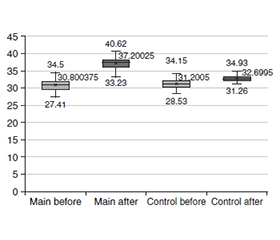
Актуальність. Цукровий діабет є однією з провідних глобальних проблем охорони здоров’я, а виразки діабетичної стопи (ВДС) належать до його найтяжчих хронічних ускладнень. ВДС характеризуються порушенням процесів репарації, ішемією, нейропатією та високим ризиком інфекційних ускладнень, що часто призводить до тривалих незагойних ран і ампутацій нижніх кінцівок. Ефективне лікування потребує комплексного підходу, зокрема своєчасного дебридменту рани, однак традиційні методи не завжди є достатньо селективними та можуть спричиняти додаткову травматизацію тканин. Останніми роками в лікування хронічних ран активно впроваджуються лазерні технології. Водночас діодні лазери залишаються недостатньо дослідженими при ішемічних формах ВДС. Мета: оцінити ефективність і безпеку лазерної вапоризації з використанням діодного лазера як ад’ювантного методу локального дебридменту при ішемічних виразках діабетичної стопи та визначити її вплив на тканинну перфузію і процеси загоєння. Матеріали та методи. У дослідження включено 120 осіб з ішемічними виразками стопи. Основна група (n = 80) отримувала стандартну терапію в поєднанні з вапоризацією діодним лазером із довжиною хвилі 1470 нм, група порівняння (n = 40) — лише стандартне лікування. Усім пацієнтам проводили доплерографію судин, визначення кісточково-плечового індексу та вимірювання транскутанної напруги кисню (TcpO2) до лікування та після 21-денного курсу терапії. Статистичну обробку виконували за допомогою стандартного програмного забезпечення; рівень статистичної значущості встановлювали при p < 0,05. Результати. До початку лікування в пацієнтів обох груп виявлено виразковi дефекти на фоні помірної або тяжкої ішемії. Після терапії в основній групі у 80 % випадків відзначено прискорення репаративних процесів, активне формування грануляцій та епітелізацію. Середній рівень TcpO2 зріс із 30,8 ± 3,0 мм рт.ст. до 37,2 ± 3,0 мм рт.ст., тоді як у групі порівняння — лише з 31,2 ± 2,0 мм рт.ст. до 32,7 ± 1,0 мм рт.ст. Середній приріст ΔTcpO2 становив 6,4 проти 1,5 мм рт.ст. відповідно (p < 0,001). Поліпшення оксигенації супроводжувалося відновленням мікроциркуляції, скороченням термінів загоєння та зменшенням тривалості госпіталізації. Висновки. Лазерна вапоризація з використанням діодного лазера є ефективним і безпечним ад’ювантним методом локального дебридменту при ішемічних ВДС. Її поєднання зі стандартною терапією сприяє вірогідному поліпшенню тканинної перфузії, прискоренню загоєння й оптимізації клінічних результатів.
Background. Diabetes mellitus is a major global health problem, and diabetic foot ulcers (DFU) represent one of its most serious chronic complications. DFUs are associated with impaired tissue repair, ischemia, neuropathy, and a high risk of infection, often leading to prolonged non-healing wounds and lower limb amputations. Effective management requires comprehensive therapy, including wound debridement; however, conventional methods may lack selectivity and cause additional tissue trauma. In recent years, laser technologies have been introduced into wound care. While CO2 and erbium-doped yttrium aluminum garnet lasers are well studied, diode lasers remain insufficiently investigated in ischemic DFU. The aim of this study was to assess the efficacy and safety of diode laser-assisted vaporization as an adjunctive local debridement method in the treatment of ischemic diabetic foot ulcers and to evaluate its effects on tissue perfusion and wound healing. Materials and methods. The study involved 120 patients with ischemic DFUs. The main group (n = 80) received standard therapy combined with laser vaporization using a 1470 nm diode laser, while the control group (n = 40) received standard treatment alone. All patients underwent Doppler ultrasound, ankle-brachial index assessment, and transcutaneous oxygen tension (TcpO2) measurement at baseline and after a 21-day treatment course. Laser parameters were individualized according to wound depth, location, and ischemia severity. Statistical analysis was performed using standard software, with significance set at p < 0.05. Results. Before treatment, patients in both groups exhibited ulcerative defects associated with moderate to severe ischemia. After therapy, accelerated reparative processes, active granulation tissue formation, and epithelialization were observed in 80 % of patients in the main group. The mean TcpO2 level increased from 30.8 ± 3.0 mmHg to 37.2 ± 3.0 mmHg in the main group, whereas in the control group, only from 31.2 ± 2.0 mmHg to 32.7 ± 1.0 mmHg. The mean increase in ΔTcpO2 was 6.4 versus 1.5 mmHg, respectively (p < 0.001). Improved tissue oxygenation was accompanied by restoration of microcirculation, shortened wound healing time, and reduced duration of hospitalization. Conclusions. Diode laser-assisted vaporization is an effective and safe adjunctive method of local debridement in ischemic DFUs. Its use in combination with standard therapy significantly improves tissue perfusion, accelerates wound healing, and enhances clinical outcomes. Further multicenter randomized studies are needed to confirm these results and optimize treatment protocols.
синдром діабетичної стопи; ішемія; транскутанна оксиметрія; мікроциркуляція; хронічна артеріальна недостатність; виразка діабетичної стопи; лазерна вапоризація
diabetic foot syndrome; ischemia; transcutaneous oximetry; microcirculation; chronic arterial insufficiency; diabetic foot ulcer; laser vaporization
Для ознакомления с полным содержанием статьи необходимо оформить подписку на журнал.
- Armstrong DG, Tan TW, Boulton AJM, Bus SA. Diabe–tic foot ulcers: a review. JAMA. 2023;330(1):62-75. doi: 10.1001/jama.2023.10578.
- Amin N, Doupis J. Diabetic foot disease: from the evaluation of the “foot at risk” to the novel diabetic ulcer treatment modalities. World J Diabetes. 2016;7(7):153-164. doi: 10.4239/wjd.v7.i7.153.
- Dawi J, Tumanyan K, Tomas K, Misakyan Y, Gargaloyan A, et al. Diabetic foot ulcers: pathophysiology, immune dysregulation, and emerging therapeutic strategies. Biomedicines. 2025;13(5):1076. doi: 10.3390/biomedicines13051076.
- Luo Y, Liu C, Li C, Jin M, Pi L, Jin Z. The incidence of lower extremity amputation and its associated risk factors in patients with diabetic foot ulcers: a meta-analysis. Int Wound J. 2024;21(7):e14931. doi: 10.1111/iwj.14931.
- McDermott K, Fang M, Boulton AJM, Selvin E, Hicks CW. Etiology, epidemiology, and disparities in the burden of diabetic foot ulcers. Diabetes Care. 2023;46(1):209-221. doi: 10.2337/dci22-0043.
- Game FL, Chipchase SY, Hubbard R, Burden RP, Jeffcoate WJ. Temporal association between the incidence of foot ulceration and the start of dialysis in diabetes mellitus. Nephrol Dial Transplant. 2006;21(11):3207-3210. doi: 10.1093/ndt/gfl427.
- Schaper NC, van Netten JJ, Apelqvist J, Bus SA, Fitridge R, et al., IWGDF Editorial Board. Practical guidelines on the prevention and management of diabetes-related foot disease (IWGDF 2023 update). Diabetes Metab Res Rev. 2024;40(3):e3657. doi: 10.1002/dmrr.3657.
- National Institute for Health and Care Excellence. Diabetic foot problems: prevention and management. London: NICE; 2023. Available from: https://www.ncbi.nlm.nih.gov/books/NBK553608/.
- Liu H, Ya-Qing X, Cai-Feng Y, Jia-Li H, Xian-Yu T. Diabetic foot wound ulcer management by laser therapy: a meta-analysis. Int Wound J. 2023;20(10):4208-4216. doi: 10.1111/iwj.14320.
- Beckmann KH, Meyer-Hamme G, Schröder S. Low level laser therapy for the treatment of diabetic foot ulcers: a critical survey. Evid Based Complement Alternat Med. 2014;2014:626127. doi: 10.1155/2014/626127.
- Huang J, Chen J, Xiong S, Huang J, Liu Z. The effect of low-level laser therapy on diabetic foot ulcers: a meta-analysis of randomised controlled trials. Int Wound J. 2021;18(6):763-776. doi: 10.1111/iwj.13577.
- Santos CMD, Rocha RBD, Hazime FA, Cardoso VS. A systematic review and meta-analysis of the effects of low-level laser therapy in the treatment of diabetic foot ulcers. Int J Low Extrem Wounds. 2021;20(3):198-207. doi: 10.1177/1534734620914439.
- Feitosa MC, Carvalho AF, Feitosa VC, Coelho IM, Oliveira RA, Arisawa EÂ. Effects of the low-level laser therapy (LLLT) in the process of healing diabetic foot ulcers. Acta Cir Bras. 2015;30(12):852-857. doi: 10.1590/S0102-865020150120000010.