
Международный эндокринологический журнал Том 22, №3, 2026
Вернуться к номеру
Вплив безпліддя на рівень білка теплового шоку 70, адропіну та орексину в сироватці крові жінок: дослідження типу «випадок — контроль»
Авторы: Noor Jassem Mohammed (1), Nagham Qasim Kadhim (1), Sabbar Rashid Lateef (2), Mohammed R. Abed Al-Joubory (3), Saif M. Hasan (4), Inaam Faisal Mohammed (5)
(1) - College of Science, Tikrit University, Iraq
(2) - Al-Iraqia University, College of Medicine, Branch of Clinical Chemistry, Iraq
(3) - Al-Imam University College, Salah Alden, Iraq
(4) - College of Health and Medical Technologies, University of Mashreq, Baghdad, Iraq
(5) - College of Medicine, University of Diyala, Iraq
Рубрики: Эндокринология
Разделы: Клинические исследования
Версия для печати
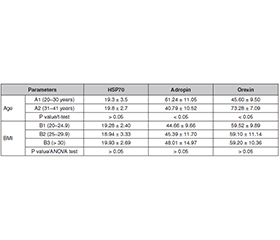
Актуальність. Безпліддя залишається серйозною проблемою репродуктивного здоров’я, яка впливає на мільйони жінок у всьому світі. Нещодавні дослідження висвітлили потенційну роль метаболічних і нейроендокринних пептидів, включаючи білок теплового шоку 70 (HSP70), адропін та орексин, у репродуктивній фізіології. Мета: оцінити рівні HSP70, адропіну та орексину в сироватці крові безплідних пацієнток порівняно з контрольною групою фертильних жінок. Матеріали та методи. Це дослідження типу «випадок — контроль», у якому взяли участь 60 пацієнток із первинним безпліддям, було проведено в Багдаді (Ірак). Тридцять здорових фертильних жінок аналогічного віку сформували контрольну групу. Вік учасниць перебував в межах від 20 до 45 років. Діагноз безпліддя було підтверджено за допомогою визначення гормонального профілю (фолікулостимулюючий та лютеїнізуючий гормон) і ультразвукового дослідження. Для оцінки рівнів HSP70, адропіну та орексину в сироватці крові використовували імуноферментний метод. Середній вміст гормонів у кожній групі порівнювали за допомогою статистичного аналізу. Результати. На відміну від контрольної групи фертильних жінок спостерігалося вірогідне підвищення (P < 0,05) рівнів фолікулостимулюючого гормону й пролактину, без суттєвої різниці (P > 0,05) у показниках лютеїнізуючого гормону. Різниця в концентрації фолікулостимулюючого гормону між групами за індексом маси тіла була вірогідною (P > 0,05), тоді як відмінностей за віком не виявлено. Результати показали, що рівень адропіну в пацієнток із первинним безпліддям вірогідно знижувався (P < 0,05). При цьому відзначалося вірогідне підвищення вмісту HSP70 та орексину порівняно з контрольною групою фертильних жінок. Спостерігалася позитивна кореляція між лютеїнізуючим та фолікулостимулюючим гормоном і пролактином, пролактином та фолікулостимулюючим гормоном, тиреотропним гормоном і пролактином, тоді як між адропіном та тиреотропним гормоном виявлено сильну негативну кореляцію. Висновки. Нижчі рівні адропіну й орексину в безплідних жінок можуть відображати порушення метаболічних та нейроендокринних шляхів, що обумовлює безпліддя. Ці біомаркери можуть слугувати потенційними індикаторами репродуктивної дисфункції.
Background. Infertility is a major reproductive health issue affecting millions of women worldwide. Recent studies have highlighted the potential roles of metabolic and neuroendocrine peptides, including heat shock protein 70 (HSP70), adropin and orexin, in reproductive physiology. This study purposed to evaluate serum levels of HSP70, adropin and orexin in infertile women compared to fertile controls. Materials and methods. This case-control research, which involved 60 women with primary infertility, was carried out in Baghdad, Iraq. Control group included 30 age-matched healthy fertile women. The age of participants ranged from 20 to 45 years. Diagnosis of infertility was confirmed through hormonal profiling (follicle-stimulating and luteinizing hormone) and ultrasonography. An enzyme-linked immunosorbent assay was used to detect the levels of serum HSP70, adropin and orexin. Mean hormone content in each group were compared using statistical analysis. Results. When compared to the control group of fertile women, there was a significant increase (P < 0.05) in follicle-stimulating hormone and prolactin levels and no significant difference (P > 0.05) in luteinizing hormone. The difference in follicle-stimulating hormone content between body mass index groups was significant (P > 0.05), and no significant difference was found in age groups. The results showed that adropin levels in the primary infertility group decreased significantly (P < 0.05), with significant increase in HSP70 and orexin when compared to control group. There was a positive correlation between luteinizing and follicle-stimulating hormone and prolactin, prolactin and follicle-stimulating hormone, thyroid-stimulating hormone and prolactin, while strong negative correlation was found between adropin and thyroid-stimulating hormone. Conclusions. Lower levels of adropin and orexin in infertile women may reflect impaired metabolic and neuroendocrine pathways contributing to infertility. These biomarkers could serve as potential indicators for reproductive dysfunction.
нейроендокринні пептиди; тиреотропний гормон; пролактин; безпліддя; репродуктивне здоров’я; адропін; орексин
neuroendocrine peptides; thyroid-stimulating hormone; prolactin; infertility; reproductive health; adropin; orexin
Для ознакомления с полным содержанием статьи необходимо оформить подписку на журнал.
- Athar F, Karmani M, Templeman NM. Metabolic hormones are integral regulators of female reproductive health and function. Biosci Rep. 2024 Jan 31;44(1):BSR20231916. doi: 10.1042/BSR20231916.
- Thatipelli RC, Parunandi Y, Nousheen S, Shenkeshi S, Fathima H, Anitha A. An observational study on causes of female infertility. International Journal of Reproduction, Contraception, Obstetrics and Gynecology. 2024;13(9):2450-2456. doi: 10.18203/2320-1770.ijrcog20242498.
- Longo M, Liuzzi F, De Carlini S, La Marca A. The role of LH in follicle development: from physiology to new clinical implications. Reprod Biol Endocrinol. 2025 Feb 10;23(Suppl 1):22. doi: 10.1186/s12958-025-01353-8.
- Behl T, Kaur I, Sehgal A, Singh S, Bhatia S, et al. The Footprint of Kynurenine Pathway in Neurodegeneration: Janus-Faced Role in Parkinson’s Disorder and Therapeutic Implications. Int J Mol Sci. 2021 Jun 23;22(13):6737. doi: 10.3390/ijms22136737.
- Brown EDL, Obeng-Gyasi B, Hall JE, Shekhar S. The Thyroid Hormone Axis and Female Reproduction. Int J Mol Sci. 2023 Jun 6;24(12):9815. doi: 10.3390/ijms24129815.
- Liu Q, Qiu Y, Jiang J, Long S, Zhu C, et al. Causal associa–tion between thyroid function and the risk of infertility: a Mendelian randomization study. Front Endocrinol (Lausanne). 2024 Oct 4;15:1425639. doi: 10.3389/fendo.2024.1425639.
- Khan AA, Sharma R, Ata F, Khalil SK, Aldien AS, et al. Systematic review of the association between thyroid disorders and hyperprolactinemia. Thyroid Res. 2025 Jan 3;18(1):1. doi: 10.1186/s13044-024-00214-7.
- Hema KR, Girish BL, Dhananjaya BS, Riyaj Ahmad Kalaburgi. Prevalence of thyroid dysfunction in women with abnormal uterine bleeding in reproductive age. Int J Reprod Contracept Obstet Gynecol. 2020;9(7):2792-2797. doi: 10.18203/2320-1770.ijrcog20202710.
- Singh MK, Han S, Ju S, Ranbhise JS, Ha J, et al. Hsp70: A Multifunctional Chaperone in Maintaining Proteostasis and Its Implications in Human Disease. Cells. 2025 Mar 29;14(7):509. doi: 10.3390/cells14070509.
- Radons J. The human HSP70 family of chaperones: where do we stand? Cell Stress Chaperones. 2016 May;21(3):379-404. doi: 10.1007/s12192-016-0676-6.
- Modrzejewska M, Zdanowska O. The Role of Heat Shock Protein 70 (HSP70) in the Pathogenesis of Ocular Diseases — Current Literature Review. J Clin Med. 2024 Jun 30;13(13):3851. doi: 10.3390/jcm13133851.
- Szyller J, Bil-Lula I. Heat Shock Proteins in Oxidative Stress and Ischemia/Reperfusion Injury and Benefits from Physical Exercises: A Review to the Current Knowledge. Oxid Med Cell Longev. 2021 Jan 31;2021:6678457. doi: 10.1155/2021/6678457.
- Maurya S, Tripathi S, Arora T, Singh A. Adropin may regulate corpus luteum formation and its function in adult mouse ovary. Hormones (Athens). 2023 Dec;22(4):725-739. doi: 10.1007/s42000-023-00476-0.
- Hasanpour-Segherlou Z, Butler AA, Candelario-Jalil E, Hoh BL. Role of the Unique Secreted Peptide Adropin in Various Phy–siological and Disease States. Biomolecules. 2024 Dec 17;14(12):1613. doi: 10.3390/biom14121613.
- Chen IW, Lin CW, Lin CN, Chen ST. Serum adropin levels as a potential biomarker for predicting diabetic kidney disease progression. Front Endocrinol (Lausanne). 2025 Feb 7;16:1511730. doi: 10.3389/fendo.2025.1511730.
- Hashimoto K. Evaluating the safety of orexin receptor antagonists on reproductive health and sexual function. Mol Psychiatry. 2025 Mar;30(3):1161-1163. doi: 10.1038/s41380-024-02858-1.
- Abdelmissih S. A Bitter Experience That Enlightens the Future: COVID-19 Neurological Affection and Perspectives on the Orexi–genic System. Cureus. 2022 Oct 28;14(10):e30788. doi: 10.7759/cureus.30788.
- Ruhrländer J, et al. The orexin system and its impact on the autonomic nervous and cardiometabolic system in post-acute sequelae of COVID-19. Biomedicines. 2025;13(3):545. doi: 10.3390/biomedi–cines13030545.
- Edinoff AN, Silverblatt NS, Vervaeke HE, Horton CC, Girma E, et al. Hyperprolactinemia, Clinical Considerations, and Infertility in Women on Antipsychotic Medications. Psychopharmacol Bull. 2021 Mar 16;51(2):131-148. doi: 10.64719/pb.4396.
- Ersoy E, Gonel A. How FSH and LH analogs affect fertility hormone results? Medicine (Baltimore). 2025 Sep 26;104(39):e44413. doi: 10.1097/MD.0000000000044413.
- Oduwole OO, Huhtaniemi IT, Misrahi M. The Roles of Luteinizing Hormone, Follicle-Stimulating Hormone and Testosterone in Spermatogenesis and Folliculogenesis Revisited. Int J Mol Sci. 2021 Nov 25;22(23):12735. doi: 10.3390/ijms222312735.
- Sisti G, Kanninen TT, Ramer I, Witkin SS. Interaction between the inducible 70-kDa heat shock protein and autophagy: effects on ferti–lity and pregnancy. Cell Stress Chaperones. 2015 Sep;20(5):753-8. doi: 10.1007/s12192-015-0609-9.
- Molvarec A, Rigó J Jr, Lázár L, Balogh K, Makó V, et al. Increased serum heat-shock protein 70 levels reflect systemic inflammation, oxidative stress and hepatocellular injury in preeclampsia. Cell Stress Chaperones. 2009 Mar;14(2):151-9. doi: 10.1007/s12192-008-0067-8.
- Didushko OM, Herych PR, Cherniavska IV, Yatsyshyn RI, Pankiv VI. Influence of the complex treatment of hypothyroidism on the leptin level in patients with primary hypothyroidism. World of Medicine and Biology. 2018;3(65):59-63. doi: 10.26724/2079-8334-2018-3-65-59-63.
- Mataji M, Frouzanian M, Kashi Z, Abdollahi A, Bahar A, Mohammadpour RA. Relationship between thyroid nodule, prolactin, and thyroid peroxidase antibody levels: A case-control study. Caspian J Intern Med. 2025 Oct 18;16(4):712-717. doi: 10.22088/cjim.16.4.712.
- Wang X, Chang X, Wang Q, Ding X, Wang J, et al. Serum Adropin Levels Are Elevated in Patients With Hyperthyroidism. Int J Endocrinol. 2024 Oct 21;2024:7144798. doi: 10.1155/2024/7144798.
- Aljwary FA, Allwsh TA. Evaluation of adropin and its relationship with metabolic disorders in polycystic ovary syndrome. Rom J Diabetes Nutr Metab Dis. 2024;31(1):93-102. doi:10.46389/rjd-2024-0093.
- Maurya S, Tripathi S, Arora T, Singh A. Adropin ameliorates reproductive dysfunctions in letrozole-induced PCOS mouse. Sci Rep. 2025 Mar 13;15(1):8659. doi: 10.1038/s41598-025-93215-x.
- Maurya S, Tripathi S, Arora T, Singh A. Adropin may re–gulate ovarian functions by improving antioxidant potential in adult mouse. J Steroid Biochem Mol Biol. 2024 Sep;242:106524. doi: 10.1016/j.jsbmb.2024.106524.
- Ye Z, Zhang C, Zhao Y. Potential effects of adropin on syste–mic metabolic and hormonal abnormalities in polycystic ovary syndrome. Reprod Biomed Online. 2021 May;42(5):1007-1014. doi: 10.1016/j.rbmo.2021.01.020.
- Cena H, Chiovato L, Nappi RE. Obesity, Polycystic Ovary Syndrome, and Infertility: A New Avenue for GLP-1 Receptor Agonists. J Clin Endocrinol Metab. 2020 Aug 1;105(8):e2695-709. doi: 10.1210/clinem/dgaa285.
- Kuliczkowska-Płaksej J, Mierzwicka A, Jończyk M, Stachowska B, Urbanovych A, Bolanowski M. Adropin in women with polycystic ovary syndrome. Endokrynol Pol. 2019;70(2):151-156. doi: 10.5603/EP.a2018.0092.
- Aggarwal G, Morley JE, Vellas B, Nguyen AD, Butler AA; MAPT/DSA Group. Low circulating adropin concentrations predict increased risk of cognitive decline in community-dwelling older adults. Geroscience. 2024 Feb;46(1):897-911. doi: 10.1007/s11357-023-00824-3.
- Butler AA, Tam CS, Stanhope KL, Wolfe BM, Ali MR, et al. Low circulating adropin concentrations with obesity and aging correlate with risk factors for metabolic disease and increase after gastric bypass surgery in humans. J Clin Endocrinol Metab. 2012 Oct;97(10):3783-91. doi: 10.1210/jc.2012-2194.
- Stevens JR, Girardet C, Zhou M, Gamie F, Aggarwal G, et al.; MAPT/IHU HealthAge Open Science group. Adropin expression reflects circadian, lipoprotein, and mitochondrial processes in human tissues. Mol Metab. 2025 Sep;99:102196. doi: 10.1016/j.molmet.2025.102196.
- Silveyra P, Cataldi NI, Lux-Lantos VA, Libertun C. Role of orexins in the hypothalamic-pituitary-ovarian relationships. Acta Physiol (Oxf). 2010 Mar;198(3):355-60. doi: 10.1111/j.1748-1716.2009.02049.x.
- Safdar M, Liang A, Rajput SA, Abbas N, Zubair M, et al. Orexin-A Regulates Follicular Growth, Proliferation, Cell Cycle and Apoptosis in Mouse Primary Granulosa Cells via the AKT/ERK Signaling Pathway. Molecules. 2021 Sep 16;26(18):5635. doi: 10.3390/molecules26185635.
- Martynska L, Wolinska-Witort E, Chmielowska M, Kalisz M, Baranowska B, Bik W. Effect of orexin A on the release of GnRH-stimu–lated gonadotrophins from cultured pituitary cells of immature and mature female rats. Neuropeptides. 2014 Aug;48(4):199-205. doi: 10.1016/j.npep.2014.05.005.