Інформація призначена тільки для фахівців сфери охорони здоров'я, осіб,
які мають вищу або середню спеціальну медичну освіту.

Підтвердіть, що Ви є фахівцем у сфері охорони здоров'я.


Коморбідний ендокринологічний пацієнт

Журнал «Травма» Том 27, №2, 2026

Вернуться к номеру

Малоінвазивна гібридна фіксація та реабілітація при переломах надп’ятково-гомілкового суглоба типу B/C за Danis-Weber у пацієнтів із цукровим діабетом: порівняльні результати

Авторы: U.V. Kuz (1), Yu.O. Divnych (2), I.M. Gudz (1), S.M. Vasyliuk (1), Ye.V. Nychvyd (1), V.I. Dubas (1), V.P. Omelchuk (1), V.S. Sulyma (1)
(1) - Ivano-Frankivsk National Medical University, Ivano-Frankivsk, Ukraine
(2) - University of Bern, Bern, Switzerland

Рубрики: Травматология и ортопедия

Разделы: Клинические исследования

Версия для печати


Резюме

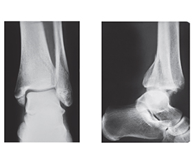
Актуальність. Хірургічне лікування пацієнтів із діабетом та переломом надп’ятково-гомілкового суглоба (НГС) є складним через високий ризик ускладнень, як-от стопа Шарко. Мета: покращити ефективність хірургічного лікування та реабілітації хворих на діабет із переломами НГС. Матеріали та методи. Дослідження провели у 31 пацієнта з переломами НГС типу B і С за класифікацією Danis-Weber. У 7 осіб із діабетом І (дослідної) групи виконано малоінвазивний (гібридний) остеосинтез за оригінальною методикою (патент № 134463); у 9 пацієнтів ІІ (порівняльної) групи без діабету, але з вираженими післятравматичними ознаками ушкодження м’яких тканин провели аналогічний остеосинтез; у 15 осіб ІІІ (контрольної) групи без хронічних патологій — остеосинтез пластиною з фіксацією дистального міжгомілкового синдесмозу позиційним шурупом. Ризик післяопераційних ускладнень у І групі прогнозували за шкалою Adelaide Fracture in the Diabetic Ankle (AFDA). Якість хірургічного лікування оцінювали за відновленням рентгеноморфометричних параметрів. Ефективність реабілітації визначали за опитувальниками American Orthopaedic Foot and Ankle Score (AOFAS) та Olerud-Molander Ankle Score (OMAS) на 4-й і 8-й тиждень після операції та через 6 місяців. ­Резу­льтати. Згідно з післяопераційними рентгенологічними даними, в усіх групах досягнуто відновлення анатомічних структур НГС (p < 0,05). Суттєвих відмінностей у наявності ознаки dime/ball між групами не виявлено ( = 0,2609; p = 0,26). Щодо функціональних результатів за шкалою AOFAS у післягострому періоді (4–8 тижнів), у пацієнтів І та ІІ груп (гібридний остеосинтез) спостерігали позитивну динаміку відновлення функції суглоба (p < 0,05), а у віддаленому періоді реабілітації (6 місяців) показники між групами статистично не відрізнялися (p = 0,1158). Натомість, за опитувальником OMAS, після гібридного остеосинтезу пацієнти мали кращі функціональні результати в післягострому періоді (4-й тиждень). На 8-му тижні в усіх групах досягнуто однаковий рівень відновлення, а на 6-му місяці у ІІ та ІІІ групах виявили вірогідно кращі результати, ніж у контрольній (р = 0,02354). Висновки. В осіб із діабетом та переломами НГС Danis-Weber типу B і С, які мають високий ризик післяопераційних ускладнень за шкалою AFDA, слід виконувати інтрамедулярний гібридний остеосинтез, що мінімізує ушкодження м’яких тканини при операції, та застосовувати індивідуальний протокол реабілітації, що надалі може запобігти виникненню стопи Шарко.

Background. Surgical treatment of ankle fractures in diabetic patients is challenging due to the high risk of complications such as Charcot foot. The purpose was to enhance the effectiveness of surgical treatment and rehabilitation for ankle fractures in patients with diabetes. Materials and methods. Research was conducted on 31 patients with Danis-Weber type B and C ankle fractures: group I (study) — 7 diabetic patients treated with minimally invasive osteosynthesis using an original technique (patent No. 134463); group II (comparative) — 9 non-diabetic patients with post-traumatic signs of soft tissue damage undergone similar osteosynthesis; group III (control) — 15 patients without chronic disease, treated with fibular plate osteosynthesis and syndesmosis fixation using a positional screw. In group I, complication risk was assessed using the Adelaide Fracture in the Diabetic Ankle (AFDA) scale. Surgical treatment quality was evaluated with X-ray morphometric parameters restoration. Rehabilitation effectiveness was measured by the American Orthopaedic Foot and Ankle Society (AOFAS) scale and Olerud-Molander Ankle Score (OMAS) at 4 and 8 weeks and 6 months after surgery. Results. Post-operative X-ray data confirm restoration of ankle anatomy in all groups (p < 0.05). No significant differences in dime/ball sign presence were observed between groups ( = 0.2609; p = 0.26). Functional assessment using the AOFAS scale showed no statistically significant difference among the groups (p = 0.1158). In terms of functional outcomes in the post-acute period (4–8 weeks) according to the AOFAS, patients of groups I and II (hybrid osteosynthesis) had positive dynamics of the joint recovery process (p < 0.05), and in the long-term rehabilitation period (6 months), the indicators did not differ between the groups (p = 0.1158). In contrast, according to the OMAS, after hybrid osteosynthesis, functional outcomes were better in the post-acute period (4 weeks). At 8 weeks, patients of all groups achieved the same level of recovery, and at 6 months, significantly better results were observed in groups II and III than in controls (p = 0.02354). Conclusions. Patients with diabetes and Danis-Weber type B or C ankle fractures who have a high AFDA-assessed risk of postoperative complications should undergo intramedullary hybrid osteosynthesis, which minimizes secondary soft-tissue damage during surgery, and follow an individualized rehabilitation protocol to further prevent the development of Charcot foot.


Ключевые слова

перелом надп’ятково-гомілкового суглоба; цукровий діабет; малоінвазивні хірургічні втручання; реабілітація

ankle fracture; diabetes mellitus; minimally invasive surgical procedures; rehabilitation


Для ознакомления с полным содержанием статьи необходимо оформить подписку на журнал.


Список литературы

1. Nash WJ, Hester T, Ha J. Current concepts and challenges in managing ankle fractures in the presence of diabetes: A systematic review of the literature. J Clin Orthop Trauma. 2021 Feb 3;17:44-53. doi: 10.1016/j.jcot.2021.01.016.
2. Amini B, Kadhm S, Trompeter A. The impact of diabetes mellitus on the management and outcome of ankle fractures. Injury. 2025 Apr;56(4):112226. doi: 10.1016/j.injury.2025.112226.
3. Goldberg EM, Polachek WS, Hynes K. Ankle Fractures in Diabetic Patients: A Critical Analysis. JBJS Rev. 2023 Mar 16;11(3). doi: 10.2106/JBJS.RVW.22.00147.
4. Van Hulten V. Bone strength, microarchitecture, and fracture risk in type 2 diabetes. Maastricht: Maastricht University, 2025. 297 p. doi: 10.26481/dis.20250213vh.
5. Hicks CW, Selvin E. Epidemiology of Peripheral Neuro-pathy and Lower Extremity Disease in Diabetes. Curr Diab Rep. 2019 Aug 27;19(10):86. doi: 10.1007/s11892-019-1212-8.
6. Tanios M, Brickman B, Cage E, Abbas K, Smith C, et al. Diabetes and Impaired Fracture Healing: A Narrative Review of Recent Literature. Curr Osteoporos Rep. 2022 Oct;20(5):229-239. doi: 10.1007/s11914-022-00740-z.
7. Marin C, Luyten FP, van der Schueren B, Kerckhofs G, Vandamme K. The Impact of Type 2 Diabetes on Bone Fracture Healing. Front Endocrinol (Lausanne). 2018 Jan 24;9:6. doi: 10.3389/fendo.2018.00006.
8. Lavery LA, Lavery DC, Green T, Hunt N, La Fontaine J, et al. Increased Risk of Nonunion and Charcot Arthropathy After Ankle Fracture in People with Diabetes. J Foot Ankle Surg. 2020 Jul-Aug;59(4):653-656. doi: 10.1053/j.jfas.2019.05.006.
9. Elgoyoushi SM, Morrah AN, Kholeif AM, Elbadry AA. Outcomes of Tibiotalocalcaneal (TTC) Nailing in Ankle Fractures in Diabetic Patients with Charcot Neuroarthropathy. Geriatr Orthop Surg Rehabil. 2025 Apr 15;16:21514593251328896. doi: 10.1177/21514593251328896.
10. Dawar A, Chundi G, Fuller Z, Ahn DB, Chopra AA, et al. Tibiotalocalcaneal Arthrodesis vs Open Reduction Internal Fixation for Trimalleolar Ankle Fractures in High-Risk Patients: A National Database Analysis. Foot Ankle Orthop. 2025 Sep 30;10(3):24730114251375027. doi: 10.1177/24730114251375027.
11. Sulyma VS, Chuzhak AV, inventors. Method of combined stable-elastic fixation of unstable injuries of the ankle joint in case of transsyndesmotic fractures of the fibula. Patent of Ukraine No. 134463, 2019 May 27.
12. Yee J, Pillai A, Ferris L. Diabetic ankle fractures: a review of the literature and an introduction to the Adelaide fracture in the diabetic ankle algorithm and score. Biomed Res Int. 2014;2014:153146. doi: 10.1155/2014/153146.
13. Rosenbaum AJ, DiPreta JA. Classifications in brief: Eichenholtz classification of Charcot arthropathy. Clin Orthop Relat Res. 2015 Mar;473(3):1168-71. doi: 10.1007/s11999-014-4059-y.
14. Lau BC, Allahabadi S, Palanca A, Oji DE. Understan–ding Radiographic Measurements Used in Foot and Ankle Surgery. J Am Acad Orthop Surg. 2022 Jan 15;30(2):e139-e154. doi: 10.5435/JAAOS-D-20-00189.
15. Sulyma VS, Filyak YO, Fedynyak MP, Chuzhak A, inventors. Rehabilitation protocol after a hybrid system of stable-elastic fixation of unstable fractures of the ankles and distal tibiofibular syndesmosis. Ukraine certificate of registration of copyright for the work No. 117011. 2023 Mar 10.
16. Bretherton CP, Claireaux HA, Achten J, Athwal A, Dutton SJ, et al. Protocol for the Weight-bearing in Ankle Fractures (WAX) trial: a multicentre prospective non-inferiority trial of early versus delayed weight-bearing after operatively mana-ged ankle fracture. BMC Musculoskelet Disord. 2021 Aug 9;22(1):672. doi: 10.1186/s12891-021-04560-7.
17. Tapaninaho K, Saarinen AJ, Ilves O, Uimonen MM, Hkkinen AH, et al. Structural validity of the foot and ankle outcome score for orthopaedic pathologies with Rasch Measurement Theory. Foot Ankle Surg. 2022 Feb;28(2):193-199. doi: 10.1016/j.fas.2021.03.005.
18. McKeown R, Parsons H, Ellard DR, Kearney RS. An evaluation of the measurement properties of the Olerud Molander Ankle Score in adults with an ankle fracture. Physiotherapy. 2021 Sep;112:1-8. doi: 10.1016/j.physio.2021.03.015.
19. Ahluwalia R, Wek C, Lewis TL, Stringfellow TD, Coffey D, et al.; HARnT Collaborative King’s College Hospital, London. Surgical Management of Complex Ankle Fractures in Patients with Diabetes: A National Retrospective Multicentre Study. J Clin Med. 2024 Jul 5;13(13):3949. doi: 10.3390/jcm13133949.
20. Gougoulias N, Oshba H, Dimitroulias A, Sakellariou A, Wee A. Ankle fractures in diabetic patients. EFORT Open Rev. 2020 Sep 10;5(8):457-463. doi: 10.1302/2058-5241.5.200025.
21. Alsheikh KA, Alrehaili HH, Alsabr AA, Alem AA, Albalawi MM, Aljaafri ZA. Outcomes of surgical management of ankle fractures in patients with diabetes. J Taibah Univ Med Sci. 2024 Jul 23;19(4):800-805. doi: 10.1016/j.jtumed.2024.07.003.
22. Cellier N. Management of hindfoot and ankle in Charcot arthropathy. EFORT Open Rev. 2025 Jun 2;10(6):327-335. doi: 10.1530/EOR-2025-0057.
23. Chen D, Liu S, Liu P, Song W. Effect of Elevated Blood Glucose on Postoperative Complications Among Diabetic Patients After Surgical Treatment of Torsional Ankle Fracture. Foot Ankle Int. 2024 Aug;45(8):839-844. doi: 10.1177/10711007241250005.
24. Lopez-Capdevila L, Rios-Ruh JM, Fortuo J, Costa AE, Santamaria-Fumas A, et al. Diabetic ankle fracture complications: a meta-analysis. Foot Ankle Surg. 2021 Oct;27(7):832-837. doi: 10.1016/j.fas.2020.10.015.
25. Manchanda K, Nakonezny P, Sathy AK, Sanders DT, Starr AJ, Wukich DK. A systematic review of ankle fracture treatment modalities in diabetic patients. J Clin Orthop Trauma. 2020 Dec 13;16:7-15. doi: 10.1016/j.jcot.2020.12.013.
26. Grote CW, Tucker W, Stumpff K, Birt MC, Horton GA. Primary Arthrodesis for Diabetic Ankle Fractures. Foot Ankle Orthop. 2020 Mar 24;5(1):2473011420908841. doi: 10.1177/2473011420908841.
27. Fadle AA, El-Adly W, Fekry MA, Osman AE, Khalifa AA. Primary arthrodesis for diabetic ankle fractures using a modified retrograde femoral intramedullary locking nail combined with lateral plating, surgical technique, and early results of a pilot study. Eur J Orthop Surg Traumatol. 2024 Jul;34(5):2549-2556. doi: 10.1007/s00590-024-03947-1.
28. Roepke WB, Haleem AM. Complications of Foot and Ankle Fractures in Diabetics. Foot Ankle Clin. 2025 Mar;30(1):173-190. doi: 10.1016/j.fcl.2024.04.004.
29. Stringfellow TD, Coffey D, Wek C, Bretherton C, Tan SP, et al.; HARnT Collaborative. Epidemiology & management of complex ankle fractures in the United Kingdom: A multicentre cohort study. Injury. 2024 Feb;55(2):111037. doi: 10.1016/j.injury.2023.111037.
30. Yawar B, Hanratty B, Asim A, Niazi AK, Khan AM. Suture-Button Versus Syndesmotic Screw Fixation of Ankle Fractures: A Comparative Retrospective Review Over One Year. Cureus. 2021 Sep 8;13(9):e17826. doi: 10.7759/cureus.17826.
31. Shimozono Y, Hurley ET, Myerson CL, Murawski CD, Kennedy JG. Suture Button Versus Syndesmotic Screw for Syndesmosis Injuries: A Meta-analysis of Randomized Controlled Trials. Am J Sports Med. 2019 Sep;47(11):2764-2771. doi: 10.1177/0363546518804804.
32. Xu B, Wang S, Tan J, Chen W, Tang KL. Comparison of Suture Button and Syndesmotic Screw for Ankle Syndesmotic Injuries: A Meta-analysis of Randomized Controlled Trials. Orthop J Sports Med. 2023 Jan 5;11(1):23259671221127665. doi: 10.1177/23259671221127665.
33. Kim GB, Park CH. Hybrid Fixation for Danis-Weber Type C Fractures With Syndesmosis Injury. Foot Ankle Int. 2021 Feb;42(2):137-144. doi: 10.1177/1071100720964799.
34. Shevate I, Salunkhe R, Pervez F, Pawar P. A Prospective Study on Fixation of Syndesmotic Ankle Injury: Tight Rope Versus Screw Fixation. Cureus. 2024 Aug 19;16(8):e67172. doi: 10.7759/cureus.67172.
35. Rder BW, Stake IK, Madsen JE, Frihagen F, Jacobsen SB, et al. Randomized trial comparing suture button with single 3.5 mm syndesmotic screw for ankle syndesmosis injury: similar results at 2 years. Acta Orthop. 2020 Dec;91(6):770-775. doi: 10.1080/17453674.2020.1818175.
36. Kong R, Viswanathan S, Razii N, Hazarika S. Surgical Outcomes of Syndesmotic Fixation of Ankle FracturesUsing Syndesmotic Screws Versus Suture Button Devices. Cureus. 2024 Jul 21;16(7):e65051. doi: 10.7759/cureus.65051.
37. Tong J, Ajrawat P, Chahal J, Daud A, Whelan DB, et al. Early Versus Delayed Weight Bearing and Mobilization After Ankle Fracture Fixation Surgery: A Systematic Review and Meta-analysis of Randomized Controlled Trials. Orthopedics. 2024 Mar-Apr;47(2):71-78. doi: 10.3928/01477447-20230804-08.
38. Lewis SR, Pritchard MW, Parker R, Searle HKC, Bec–kenkamp PR, et al. Rehabilitation for ankle fractures in adults. Cochrane Database Syst Rev. 2024 Sep 23;9(9):CD005595. doi: 10.1002/14651858.CD005595.pub4.
39. Khojaly R, Rowan FE, Hassan M, Hanna S, Mac Niocail R. Weight-bearing Allowed Following Internal Fixation of Ankle Fractures, a Systematic Literature Review and Meta-Analysis. Foot Ankle Int. 2022 Sep;43(9):1143-1156. doi: 10.1177/10711007221102142.
40. Bazarov I, Peace RA, Lagaay PM, Patel SB, Lyon LL, Schuberth JM. Early Protected Weightbearing After Ankle Fractures in Patients With Diabetes Mellitus. J Foot Ankle Surg. 2017 Jan-Feb;56(1):30-33. doi: 10.1053/j.jfas.2016.09.010.
41. Lorente A, Ganda A, Mariscal G, Palacios P, Lorente R. Quality of life and complications in elderly patients after pronation rotation type III ankle fractures treated with a cast and early weight-bearing. BMC Musculoskelet Disord. 2021 Oct 14;22(1):878. doi: 10.1186/s12891-021-04745-0.

Вернуться к номеру