Інформація призначена тільки для фахівців сфери охорони здоров'я, осіб,
які мають вищу або середню спеціальну медичну освіту.

Підтвердіть, що Ви є фахівцем у сфері охорони здоров'я.


Коморбідний ендокринологічний пацієнт

Журнал «Травма» Том 27, №2, 2026

Вернуться к номеру

Біомеханічні передумови тендинопатії власної зв’язки надколінка після артроскопії колінного суглоба

Авторы: Герасименко А.С., Юрик О.Є., Герасименко С.І., Бабко А.М., Полулях Д.М., Громадський В.В., Поліщук О.С.
Державна установа «Національний інститут травматології та ортопедії НАМН України», м. Київ, Україна

Рубрики: Травматология и ортопедия

Разделы: Клинические исследования

Версия для печати


Резюме

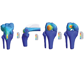
Актуальність. Тендинопатія власної зв’язки надколінка — поширена проблема у пацієнтів після артроскопічних втручань, що супроводжується хронічним болем і обмеженнями у функціональності колінного суглоба. Мета: провести комплексний біомеханічний аналіз впливу гіпотрофії квадрицепса стегна на біомеханіку колінного суглоба та механізми розвитку тендинопатії надколінка за допомогою математичного моделювання. Матеріали та методи. Моделювання кісток, зв’язок, хрящів виконувалося в програмі SolidWorks 2023 (Dassaul systemes, Франція), 3Ds Max 2025 (Autodesk Іnc, США). Дослідження методом скінченних елементів (FEM) — Simsolid 2023 (Altair Engineering, США). Результати та висновки. Було розроблено та успішно застосовано чисельну математичну модель колінного суглоба, яка дозволила оцінити розподіл напружень і контактних тисків у колінному суглобі за умов контрольованого навантаження. Ця модель враховує морфометричні параметри та фізико-механічні властивості тканин, а також дозволяє моделювати сценарії з гіпотрофією квадрицепса стегна. Моделювання біомеханіки здорового колінного суглоба при кутах згинання 30°, 60° та 90° показало закономірне зростання локальних піків напружень і контактних сил зі збільшенням кута згинання. Максимальні напруження зосереджувалися в зонах контакту патели зі стегновою кісткою та біля переходів геометрії. При 90° згинання пателофеморальна контактна сила становила 2058 Н, а напруження зв’язки досягало 3,28 МПа. Виявлено, що гіпотрофія квадрицепса стегна призводить до значного перерозподілу навантажень у пателофеморальному суглобі та сухожилку надколінка. Спостерігається виражена латералізація контактної зони та підвищення локальних піків напружень у пателофеморальному сегменті. При 30° згинання з гіпотрофією латеральна сила (621,4 Н) переважала над медіальною (221 Н), а напруження у зв’язці становило 2,4 МПа. При гіпотрофії квадрицепса спостерігається зростання напружень у зонах прикріплення сухожилка та зв’язкового апарату. Зокрема, при 60° згинання з гіпотрофією напруження у зв’язці становило 6,8 МПа, а локальні піки в пателофеморальному відділі досягали 13,116 МПа. Це свідчить про підвищений ризик перевантаження латеральних структур та сухожилка. Гіпотрофія квадрицепса стегна створює умови для збільшення моментів, що формують ексцентричне навантаження (наприклад, 9,4 Н·м при 30° згинання). Ці моменти вказують на формування ротаційного та згинального ефекту, здатного посилювати локальні напруження та сприяти мікротравматизації зв’язки.

Background. Patellar tendinopathy is a common problem in patients after arthroscopic surgery, accompanied by chronic pain and limitations in knee joint function. The purpose of the study was to conduct a comprehensive biomechanical analysis of the effect of quadriceps hypotrophy on the biomechanics of the knee joint and the mechanisms of patellar tendinopathy development using mathe-matical modelling. Materials and methods. Modelling of bones, li­gaments, and cartilages was performed using SolidWorks 2023 (Dassault Systmes, France) and 3ds Max 2025 (Autodesk Inc, USA). Finite element method research was performed using SimSolid 2023 (Altair Engineering, USA). Results and conclusions. A numerical mathematical model of the knee joint was developed and successfully applied, which made it possible to evaluate the distribution of stresses and contact pressures in the knee joint under controlled load. This model considers the morphometric parameters, physical and mechanical properties of tissues, and also allows modelling scenarios with quadriceps hypotrophy. Modelling the biomechanics of a healthy knee joint at flexion angles of 30, 60 and 90° showed a regular increase in local stress peaks and contact forces with an increase in flexion angle. The maximum stresses were concentrated in the areas of contact between the patella and the femur and near the geo­metry transitions. At 90° flexion, the шpatellofemoral contact force was 2058 N, and the ligament stress reached 3.28 MPa. It was found that quadriceps hypotrophy leads to a significant redistribution of loads in the patellofemoral joint and patellar tendon. There are a pronounced lateralisation of the contact zone and an increase in local stress peaks in the patellofemoral segment. At 30° flexion with hypotrophy, the la­teral force (621.4 N) prevailed over the medial force (221 N), and the stress in the ligament was 2.4 MPa. With quadriceps hypotrophy, there is an increase in stress in the areas of attachment of the tendon and ligamentous apparatus. In particular, at 60° flexion with hypotrophy, the stress in the ligament was 6.8 MPa, and local peaks in the patellofemoral region reached 13.116 MPa. This indicates an increased risk of overload of the lateral structures and tendon. Quadriceps hypotrophy creates conditions for an increase in moments that form an eccentric load (for example, 9.4 Nm at 30° flexion). These moments indicate the formation of a rotational and flexion effect that can increase local stresses and contribute to microtrauma of the ligament.


Ключевые слова

тендинопатія надколінка; артроскопія колінного суглоба; реабілітація; роботизовані ортези; синдром «переднього болю» колінного суглоба

patellar tendinopathy; knee arthroscopy; rehabilitation; robotic orthoses; anterior knee pain syndrome


Для ознакомления с полным содержанием статьи необходимо оформить подписку на журнал.


Список литературы

1. Milczarek, J., Szymczuk, B., Iwan, M., Trojanowski, D., Adamiec, K., … & Nitka, K. (2024). Review of the Literature on Patellar Tendinopathy: Current State of Knowledge. Quality in Sport, 20, 51433. https://doi.org/10.12775/qs.2024.20.51433. 
2. Nutarelli, S., Lodi, C.M.T. da, Cook, J., Deabate, L., & Filardo, G. (2023). Epidemiology of Patellar Tendinopathy in Athletes and the General Population: A Systematic Review and Meta-analysis [Review of Epidemiology of Patellar Tendinopathy in Athletes and the General Population: A Systematic Review and Meta-analysis]. Orthopaedic Journal of Sports Medicine, 11(6). SAGE Publishing. https://doi.org/10.1177/23259671231173659. 
3. Tayfur, A., Haque, A., Salles-Neto, J.I., Malliaras, P., Screen, H.R.C., & Morrissey, D. (2021). Are Landing Patterns in Jumping Athletes Associated with Patellar Tendinopathy? A Systematic Review with Evidence Gap Map and Meta-analysis [Review of Are Landing Patterns in Jumping Athletes Associated with Patellar Tendinopathy? A Systematic Review with Evidence Gap Map and Meta-analysis]. Sports Medicine, 52(1), 123. Springer Science+Business Media. https://doi.org/10.1007/s40279-021-01550-6. 
4. Garcia, S., Delattre, N., Berton, ., & Rao, G. (2022). Patellar Tendon Force Differs Depending on Jump-Landing Tasks and Estimation Methods. Applied Sciences, 12(1), 488. https://doi.org/10.3390/app12010488. 
5. Feng, R., Best, T.M., Wang, L., Gao, W., Liu, H., & Yu, B. (2022). Knee Movement Characteristics of Basketball Pla–yers in Landing Tasks Before Onset of Patellar Tendinopathy: A Prospective Study. Frontiers in Sports and Active Living, 4. https://doi.org/10.3389/fspor.2022.847945. 
6. Mohamed, S.H., Mohamed, G.E., Mosaad, D.M., Abdelazeim, A.S., Genedy, A., & Elhafez, H.M. (2021). Effect of ischaemic compression versus kinesiotape on patellar tendinitis. Physiotherapy Quarterly, 29(1), 47. https://doi.org/10.5114/pq.2020.96425. 
7. Thomas, A.C. (2010). Muscle Dysfunction Associated With ACL Injury and Reconstruction. Deep Blue (University of Michigan). https://hdl.handle.net/2027.42/78807. 
8. Chavan, S., & Wabale, R.N. (2016). Reviewing morpho–logy of Quadriceps femoris muscle. Journal of Morphological Sciences, 33(2), 112. https://doi.org/10.4322/jms.053513. 
9. Winkler, T., Bell, L.G., Bender, A., Trepczynski, A., Duda, G.N., Baur, A.J.D., & Damm, P. (2023). Periarticular muscle status affects in vivo tibio-femoral joint loads after total knee arthroplasty. Frontiers in Bioengineering and Biotechno–logy, 11. https://doi.org/10.3389/fbioe.2023.1075357. 
10. Mebarki, S., Jourdan, F., Canovas, F., Malachanne, E., & Dagneaux, L. (2022). Validation of a novel finite-element model for evaluating patellofemoral forces and stress du–ring squatting after posterior-stabilized total knee arthroplasty. Orthopaedics & Traumatology Surgery & Research, 109(5), 103519. https://doi.org/10.1016/j.otsr.2022.103519 
11. Wang, X., Duan, L., Liu, H., Ge, H., Dong, Z., … & Wang, F. (2023). The influence of varus and valgus deviation on the contact area of patellofemoral joint in healthy knees. BMC Musculoskeletal Disorders, 24(1). https://doi.org/10.1186/s12891-023-06976-9. 
12. Wang, X., Liu, H., Dong, Z., Chen, X., Xu, C., … & Wang, F. (2023). Contact area and pressure changes of patellofemoral joint during stair ascent and stair descent. BMC Musculoskeletal Disorders, 24(1). https://doi.org/10.1186/s12891-023-06882-0. 
13. Rood, A., Hannink, G., Lenting, A., Groenen, K.H.J., Koter, S., Verdonschot, N., & Kampen, A. van (2015). Patellofemoral Pressure Changes After Static and Dynamic Medial Patellofemoral Ligament Reconstructions. The American Journal of Sports Medicine, 43(10), 2538. https://doi.org/10.1177/0363546515594447. 
14. Pal, S., Besier, T.F., Gold, G.E., Fredericson, M., Delp, S.L., & Beaupr, G.S. (2018). Patellofemoral cartilage stresses are most sensitive to variations in vastus medialis muscle forces. Computer Methods in Biomechanics & Biomedical Engineering, 22(2), 206. https://doi.org/10.1080/10255842.2018.1544629.
15. Stephen, J.M., Alva, A., Lumpaopong, P., Williams, A., & Amis, A.A. (2018). A cadaveric model to evaluate the effect of unloading the medial quadriceps on patellar tracking and patellofemoral joint pressure and stability. Journal of Experimental Orthopaedics, 5(1). https://doi.org/10.1186/s40634-018-0150-8. 
16. Owusu-Akyaw, K., Heckelman, L.N., Cutcliffe, H.C., Sutter, E.G., Englander, Z.A., … & DeFrate, L.E. (2017). A comparison of patellofemoral cartilage morphology and deformation in anterior cruciate ligament deficient versus uninjured knees. Journal of Biomechanics, 67, 78. https://doi.org/10.1016/j.jbiomech.2017.11.019.
17. Claudon, B. (2010). Kinetic gait adaptation in patients with symptomatic patellofemoral syndrome: reversibility under rehabilitation program. HAL (Le Centre Pour La Communication Scientifique Directe). https://hal.univ-lorraine.fr/hal-01733971. 
18. Wheatley, M.G.A., Rainbow, M.J., & Clouthier, A.L. (2020). Patellofemoral Mechanics: a Review of Pathomechanics and Research Approaches [Review of Patellofemoral Mecha–nics: a Review of Pathomechanics and Research Approaches]. Current Reviews in Musculoskeletal Medicine, 13(3), 326. Springer Science+Business Media. https://doi.org/10.1007/s12178-020-09626-y.
19. Sandercock, T.G., Wei, Q., Dhaher, Y.Y., Pai, D.K., & Tresch, M.C. (2018). Vastus lateralis and vastus medialis produce distinct mediolateral forces on the patella but similar forces on the tibia in the rat. Journal of Biomechanics, 81, 45. https://doi.org/10.1016/j.jbiomech.2018.09.007.
20. Stefanik, J.J., Guermazi, A., Zhu, Y., Zumwalt, A., Gross, K.D., … & Felson, D.T. (2011). Quadriceps weakness, patella alta, and structural features of patellofemoral osteoarthritis. Arthritis Care & Research, 63(10), 1391. https://doi.org/10.1002/acr.20528.
21. Lin, F., Wilson, N.A., Makhsous, M., Press, J.M., Koh, J.L., Nuber, G.W., & Zhang, L. (2009). In vivo patellar trac–king induced by individual quadriceps components in individuals with patellofemoral pain. Journal of Biomechanics, 43(2), 235. https://doi.org/10.1016/j.jbiomech.2009.08.043. 

Вернуться к номеру