
Архив офтальмологии Украины Том 14, №1, 2026
Вернуться к номеру
Віддалені результати ексимерлазерних втручань в комплексному лікуванні кератоконуса: 2 роки спостереження
Авторы: Могілевський С.Ю. (1), Афанасьєв І.В. (1, 2), Денисюк Л.І. (1)
(1) - Національний університет охорони здоров’я України імені П.Л. Шупика, м. Київ, Україна
(2) - Офтальмологічний центр ТОВ «Ексімер Одеса», м. Одеса, Україна
Рубрики: Офтальмология
Разделы: Клинические исследования
Версия для печати
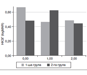
Актуальність. Кератоконус є поширеним прогресуючим захворюванням рогівки, яке призводить до зниження зорових функцій та якості життя пацієнтів. За даними метааналізу, його поширеність становить 138 на 100 000 населення. Сучасні підходи до лікування спрямовані не лише на стабілізацію ектатичного процесу за допомогою рогівкового крослінкінгу (CXL), а й на поліпшення функціональних результатів шляхом поєднання з ексимерлазерними втручаннями. Однак питання довготривалої ефективності, стабільності результатів та впливу таких комбінованих методів на зорові функції залишаються недостатньо вивченими. Мета: дослідити ефективність різних методів ексимерлазерних втручань у комплексному лікуванні кератоконуса при терміні спостереження 2 роки. Матеріали та методи. У дослідження було включено 58 пацієнтів (64 ока). З них було 40 чоловіків (69 %) і 18 жінок (31 %), віком від 20 до 40 років, що становили дві групи залежно від методу втручання. Пацієнтам 1-ї групи виконано фоторефракційну кератектомію під контролем топографії з конвенційним (за Дрезденським протоколом) крослінкінгом (TG PRK з CXL). Пацієнтам 2-ї групи виконано трансепітеліальну фототерапевтичну кератектомію під контролем топографії (без корекції рефракційного компонента) з конвенційним (за Дрезденським протоколом) крослінкінгом (TG t-PTK з CXL). Результати. TG-PRK з CXL та TG t-PTK з CXL продемонстрували задовільний профіль безпеки та були ефективними в поліпшенні гостроти зору у віддаленому періоді спостереження. Частота більшості післяопераційних ускладнень статистично не відрізнялася між групами. Водночас через 1 рік після втручання у 1-й групі спостереження відзначалася вірогідно вища частота помутніння рогівки порівняно з 2-ю групою, однак зазначені зміни були транзиторними і регресували протягом 2 років спостереження. Частота прогресування кератоконуса відзначалась тільки в 1-й групі, але була статистично незначущою. Максимально коригована гострота зору (МКГЗ) вірогідно підвищувалася після лікування, причому у 2-й групі отриманий ефект був більш стабільним у віддаленому періоді спостереження. Некоригована гострота зору (НКГЗ) характеризувалася тимчасовим погіршенням через 1 рік із подальшим відновленням до вихідного рівня через 2 роки. Міжгрупове порівняння не виявило статистично значущих відмінностей за показниками НКГЗ та МКГЗ на жодному з етапів спостереження. Висновки. У результаті проведених нами досліджень встановлено, що TG PRK з CXL та TG t-PTK з CXL продемонстрували задовільний профіль безпеки у віддаленому періоді спостереження. Частота більшості ускладнень не відрізнялася між групами. Водночас через 1 рік у 1-й групі спостереження відзначалося вірогідно вище помутніння рогівки (23,5 проти 3 %, p = 0,029), яке було транзиторним та регресувало через 2 роки після втручання. Низька та статистично незначуща частота прогресування кератоконуса в 1-й групі, а також відсутність міжгрупових відмінностей підтверджують ефективність комбінованого підходу в стабілізації ектатичного процесу при терміні спостереження 2 роки. Проведене лікування забезпечувало поліпшення зорових функцій в обох групах спостереження. МКГЗ вірогідно підвищувалася, причому у 2-й групі ефект був більш стабільним у віддаленому періоді. НКГЗ демонструвала тимчасове погіршення через 1 рік із подальшим відновленням до вихідного рівня через 2 роки. Статистично значущих відмінностей між групами за показниками НКГЗ та МКГЗ не виявлено. Дослідження нових факторів ризику, етіологічних та патогенетичних чинників кератоконуса дозволять підвищити ефективність лікування цієї складної очної патології.
Background. Keratoconus is a common progressive corneal disorder that leads to a decline in visual function and quality of life. According to meta-analytical data, its prevalence is approximately 138 cases per 100,000 population. Current treatment strategies are aimed not only at stabilizing the ectatic process using corneal cross-linking (CXL), but also at improving functional outcomes through its combination with excimer laser procedures. However, the issues of long-term efficacy, stability of outcomes, and the impact of such combined approaches on visual function remain insufficiently investigated. The purpose was to evaluate the effectiveness of different excimer laser techniques as part of combined treatment for keratoconus over a 2-year follow-up period. Materials and methods. The study included 58 patients (64 eyes), comprising 40 men (69 %) and 18 women (31 %) aged 20 to 40 years. Participants were divided into two groups based on the intervention method. Group 1 underwent topography-guided photorefractive keratectomy combined with conventional (Dresden) crosslinking (TG-PRK with CXL). Group 2 included patients who underwent topography-guided transepithelial phototherapeutic keratectomy (without correction of the refractive component) with conventional (Dresden) crosslinking (TG t-PTK with CXL). Results. TG-PRK with CXL and TG t-PTK with CXL demonstrated a favorable safety profile and were effective in improving visual acuity over a long-term follow-up period. The incidence of most postoperative complications did not differ significantly between the groups. However, at 1 year postoperatively, the first group showed a significantly higher rate of corneal haze compared to the second group; these changes were transient and regressed within a 2-year follow-up period. Keratoconus progression was observed only in the first group, but the difference was not statistically significant. Best corrected visual acuity (BCVA) improved significantly after treatment, with a more stable effect observed in the second group over the long term. Uncorrected visual acuity (UCVA) demonstrated a temporary deterioration at 1 year, followed by a return to baseline values at 2 years. No statistically significant intergroup differences were found in UCVA or BCVA at any follow-up time point. Conclusions. The present study demonstrated that TG-PRK with CXL and TG t-PTK with CXL provided a favorable safety profile over a long-term follow-up period. The incidence of most complications did not differ significantly between the groups. However, at 1 year, a significantly higher rate of corneal haze was observed in the first group (23.5 vs 3 %, p = 0.029); these changes were transient and regressed within 2 years after the intervention. The low and statistically non-significant rate of keratoconus progression in the first group, along with the absence of intergroup differences, confirms the effectiveness of combined approach in stabilizing the ectatic process over a 2-year follow-up period. The applied treatment resulted in improved visual function in both groups. BCVA increased significantly, with a more stable long-term effect observed in the second group. UCVA showed a temporary deterioration at 1 year, followed by recovery to baseline values at 2 years. No statistically significant differences between the groups were found for UCVA or BCVA. Further investigation of novel risk factors, as well as etiological and pathogenetic mechanisms of keratoconus, may contribute to improving the effectiveness of treatment for this complex ocular disease.
кератоконус; фоторефракційна кератектомія під контролем топографії; фототерапевтична кератектомія під контролем топографії; комбіноване лікування
keratoconus; topography-guided photorefractive keratectomy; topography-guided phototherapeutic keratectomy; combined treatment
Для ознакомления с полным содержанием статьи необходимо оформить подписку на журнал.
- Niazi S., Jiménez-García M., Findl O., Gatzioufas Z., Doroodgar F. et al. Keratoconus diagnosis: from fundamentals to artificial intelligence: a systematic narrative review. Diagnostics. 2023;13:2715. https://doi.org/10.3390/diagnostics13162715.
- Lucas S.E.M., Burdon K.P. Genetic and environmental risk factors for keratoconus. Annual Review of Vision Science. 2020 Sep 15;6:25-46. doi: 10.1146/annurev-vision-121219-081723.
- Erdinest N., London N., Landau D., Barbara R., Barbara A., Naroo S.A. Higher order aberrations in keratoconus. International Ophthalmology. 2024;44(1):172. https://doi.org/10.1007/s10792-024-03118-5.
- Niazi S., Gatzioufas Z., Doroodgar F., Shahriari M.H., Javadi M.A. Keratoconus: exploring fundamentals and future perspectives — a comprehensive systematic review. Therapeutic Advances in Ophthalmology. 2024;16. https://doi.org/10.1177/25158414241232258.
- Papachristoforou N., Ueno A., Ledwos K., Bartuś J., Nowiń–ska A., Karska-Basta I. A review of keratoconus cross-linking treatment methods. Journal of Clinical Medicine. 2025;14:1702. https://doi.org/10.3390/jcm14051702.
- Kumar K.K., Nataraj S., Maganty V., Porwal K.H., Gangasagara S.B. Combined topography-guided photorefractive keratectomy and corneal collagen crosslinking. Indian Journal of Ophthalmology. 2024;72(Suppl 4):S639-S644. https://doi.org/10.4103/IJO.IJO_791_23.
- Gil P., Gil J.Q., Dias M., Cortinhal T., Alves N. et al. Safety and efficacy of corneal cross-linking combined with phototherapeutic keratectomy with or without topography-guided photorefractive keratectomy. Cornea. 2025 Jun 10. https://doi.org/10.1097/ICO.0000000000003921.
- Kankariya V.P., Dube A.B., Grentzelos M.A., Kontada–kis G.A., Diakonis V.F. et al. Corneal cross-linking combined with refractive surgery for keratoconus: CXL plus. Indian Journal of Ophthalmology. 2020;68(12):2757-2772. https://doi.org/10.4103/ijo.IJO_1841_20.
- Jaruseviciene R., Tamuleviciute R., Galgauskas S. Corneal cross-linking in keratoconus: comparative analysis of standard, accelerated and transepithelial protocols. Journal of Clinical Medicine. 2026;15:490. https://doi.org/10.3390/jcm15020490.
- Афанасьєв І.В. Ефективність різних методів ексимерлазерних втручань у комплексному лікуванні кератоконуса. Архів офтальмології України. 2025;13(1):19-27. https://doi.org/10.22141/2309-8147.13.1.2025.401.
- Gijs M., Adelaar T.I., Vergouwen D.P.C., Visser N., Dick–man M.M., Ollivier R.C.I. et al. Tear fluid inflammatory proteome analysis highlights similarities between keratoconus and allergic conjunctivitis. Investigative Ophthalmology & Visual Science. 2023;64(15):9. https://doi.org/10.1167/iovs.64.15.9.
- Moura G.S., Santos A., Cenedeze M.A., Hiyane M.I., Camara N.O.S., Barbosa de Sousa L. et al. Increased lacrimal inflammatory mediators in patients with keratoconus. Molecular Vision. 2021;27:656-665.
- Krok M., Wróblewska-Czajka E., Łach-Wojnarowicz O., Bronikowska J., Czuba Z.P. et al. Analysis of cytokine and chemokine level in tear film in keratoconus patients before and after corneal cross-linking. International Journal of Molecular Sciences. 2024;25:1052. https://doi.org/10.3390/ijms25021052.
- D’Souza S., Nair A.P., Sahu G.R. et al. Keratoconus patients exhibit a distinct ocular surface immune cell and inflammatory profile. Scientific Reports. 2021;11:20891. https://doi.org/10.1038/s41598-021-99805-9.
- Cappellani F., Capobianco M., Visalli F., Mazzotta C., D’Esposito F., Tognetto D. et al. The genetic and epigenetic architecture of keratoconus. Genes. 2026;17(1):66. https://doi.org/10.3390/genes17010066.
- Alqudah N., Al-Azzam N., El Taani L., Sharayah A., Al Qudah M., Mhedat K. et al. Assessment of serum cytokine levels in keratoconus patients. Journal of Clinical Medicine. 2025;14:3179. https://doi.org/10.3390/jcm14093179.
- Rong S.S., Ma S.T.U., Yu X.T. Genetic associations for ke–ratoconus: a systematic review and meta-analysis. Scientific Reports. 2017;7:4620. https://doi.org/10.1038/s41598-017-04393-2.
- Chen J., Jia Y., Qi X. Mendelian randomization and genetic analyses reveal causal roles of immune cells and inflammatory proteins in keratoconus. Scientific Reports. 2025;15:25846. https://doi.org/10.1038/s41598-025-10759-8.
- McComish B.J., Sahebjada S., Bykhovskaya Y. et al. Association of genetic variation with keratoconus. JAMA Ophthalmology. 2020;138(2):174-181. https://doi.org/10.1001/jamaophthalmol.2019.5293.
- Cerván-Martín M., Higueras-Serrano I., González-Muñoz S. et al. Comprehensive evaluation of the genetic basis of keratoconus. Investigative Ophthalmology & Visual Science. 2024;65(12):32. https://doi.org/10.1167/iovs.65.12.32.
- Amsler M. Kératocône classique et kératocône fruste. Annales d’Oculistique. 1947;180:112.
- Ocular Surface Disease Index (OSDI). Administration and scoring manual. Allergan. 2004.
- Fantes F.E., Hanna K.D., Waring G.O., Pouliquen Y., Thompson K.P., Savoldelli M. Wound healing after excimer laser ke–ratectomy. Archives of Ophthalmology. 1990;108:665-675. https://doi.org/10.1001/archopht.1990.01070070051034.
- Gomes J.A.P., Tan D., Rapuano C.J. Global consensus on keratoconus. Cornea. 2018;37(4):359-369. https://doi.org/10.1097/ICO.0000000000001507.
- Hersh P.S., Stulting R.D., Muller D. Corneal collagen cross-linking for keratoconus. Journal of Cataract and Refractive Surgery. 2018;44(5):623-633. https://doi.org/10.1016/j.jcrs.2018.03.023.
- Mazzotta C., Traversi C., Baiocchi S. Corneal haze after PRK and CXL. Eye and Vision. 2020;7:1-9. https://doi.org/10.1186/s40662-020-00188-7.
- Kymionis G.D., Grentzelos M.A., Plaka A.D. Long-term results of combined PRK and CXL. Survey of Ophthalmology. 2023;68(2):123-135. https://doi.org/10.1016/j.survophthal.2022.06.002.
- Kanellopoulos A.J. Ten-year outcomes of keratoconus management with the Athens protocol. Journal of Refractive Surgery. 2019;35(8):478-483. https://doi.org/10.3928/1081597X-20190627-01.
- Shetty R., Dupps W.J., Roberts C.J. Biomechanics of keratoconus. Progress in Retinal and Eye Research. 2022;89:101038. https://doi.org/10.1016/j.preteyeres.2021.101038.
- Wu T, Wang Y, Li Y, Li Y, Jiang X, Li X. The impact of corneal higher-order aberrations on dynamic visual acuity post cataract surgery. Front Neurosci. 2024 May 13;18:1321423. doi: 10.3389/fnins.2024.1321423.
- Zhang H., Cantó-Cerdán M., Félix-Espinar B., Alió Del Barrio J.L. Efficacy of customized photorefractive keratectomy with cross-linking versus cross-linking alone. American Journal of Ophthalmology. 2025;274:9-23. https://doi.org/10.1016/j.ajo.2025.02.038.
- Kanellopoulos A.J. Topography-guided photorefractive kera–tectomy combined with corneal cross-linking. Journal of Refractive Surgery. 2019;35(7):478-483. https://doi.org/10.3928/1081597X-20190617-01.