
Архив офтальмологии Украины Том 14, №1, 2026
Вернуться к номеру
Роль оптичної когерентної томографії в комплексному обстеженні пацієнтів з перенесеним інсультом
Авторы: Павленко Д.О., Жабоєдов Д.Г.
Національний медичний університет імені О.О. Богомольця, м. Київ, Україна
Рубрики: Офтальмология
Разделы: Клинические исследования
Версия для печати
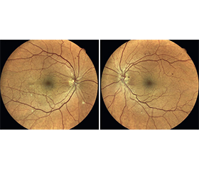
Актуальність. Сучасні високотехнологічні методи оптичної когерентної томографії (ОКТ) та ОКТ в режимі ангіографії дозволяють визначити, кількісно оцінити та відстежити з часом нейродегенеративні зміни сітківки й зорового нерва після інсульту. Минулі дослідження вказують на сильний зв’язок між структурними порушеннями у сітківці та в головному мозку, а зміни в судинах сітківки можуть передбачити ризик інсульту в пацієнта. Мета: визначити стан сітківки після інсульту та навести клінічний випадок пацієнта з постінсультною геміанопсією. Матеріали та методи. Для прикладу оцінки стану сітківки та зорового нерва із застосуванням ОКТ та ОКТ-ангіографії в комплексному обстеженні пацієнтів з перенесеним інсультом наводимо клінічний випадок пацієнта з постінсультною геміанопсією. Пацієнту було проведено такі офтальмологічні обстеження: візіометрія, авторефкератометрія, тонометрія, біомікроскопія, офтальмоскопія, фундус-фотографування, статична автоматизована периметрія, ОКТ та ОКТ-ангіографія дисків зорових нервів і макулярної ділянки сітківки. Спостереження в динаміці відбувалося на 2, 3, 4-му тижні та 3, 6, 12-му місяці після інсульту. Результати. Після інсульту виявлено поступове зниження щільності судин поверхневого та глибокого судинних комплексів макули та перипапілярних капілярів переважно всередині диска зорового нерва при відносно стабільних функціональних показниках периметрії. У пацієнта не відмічається стоншення гангліонарного комплексу сітківки протягом періоду спостереження. В оці зі сторони інсульту відмічалось стоншення шару нервових волокон сітківки. Висновки. Застосування ОКТ та ОКТ-ангіографії дозволяє швидко та неінвазивно зафіксувати наявність і відстежити нейродегенеративні та мікроваскулярні зміни сітківки у пацієнтів з перенесеним інсультом, які не є статичними і можуть бути більш виражені в оці зі сторони інсульту.
Background. The modern high-tech methods of optical coherence tomography (OCT) and OCT angiography allow us to identify, quantify and track over time neurodegenerative changes in the retina and optic nerve after stroke. Previous studies indicate a strong relationship between structural abnormalities in the retina and the brain, and changes in retinal vessels can predict the risk of stroke in a patient. The purpose was to investigate the retinal state after stroke and present a clinical case of a patient with post-stroke hemianopsia. Materials and methods. As an example of assessing the state of the retina and optic nerve using OCT and OCT angiography in a comprehensive examination of people with stroke, we present a clinical case of the patient with stroke. The patient underwent the following ophthalmological examinations: visual acuity test, autorefkeratometry, tonometry, biomicroscopy, ophthalmoscopy, fundus photography, static automated perimetry, OCT and OCT angiography of the optic nerve discs and the macular area of the retina. Prospective observation took place at weeks 2, 3, 4 and 3, 6, and 12 months after stroke. Results. After stroke, a gradual decrease in the vascular density of the superficial and deep vascular complexes of the macula and peripapillary capillaries was detected mainly inside the optic nerve disc, with relatively stable functional parameters on automated perimetry. The patient did not have thinning of the ganglion layer during the observation period. In the eye on the side of stroke, a thinning of the retinal nerve fiber layer was noted. Conclusions. The use of OCT and OCT angiography allows for rapid and noninvasive detection and monitoring of neurodegenerative and microvascular retinal changes in stroke patients, which are not static and may be more pronounced in the eye on the side of stroke.
оптична когерентна томографія; оптична когерентна томографія-ангіографія; мікроциркуляція; інсульт; сітківка; зоровий нерв
optical coherence tomography; optical coherence tomography angiography; microcirculation; stroke; retina; optic nerve
Для ознакомления с полным содержанием статьи необходимо оформить подписку на журнал.
- Rowe FJ, Hepworth LR, Howard C, et al. Impact of visual impairment following stroke (IVIS study): a prospective clinical profile of central and peripheral visual deficits, eye movement abnormalities and visual perceptual deficits. Disabil Rehabil. 2022;44:3139-3153. doi: 10.1080/09638288.2020.1859631.
- Johnson CA, Wall M, Thompson HS. A History of Perimetry and Visual Field Testing. Optom Vis Sci. 2011;88:E8-E15. doi: 10.1097/OPX.0b013e3182004c3b.
- London A, Benhar I, Schwartz M. The retina as a window to the brain — from eye research to CNS disorders. Nat Rev Neurol. 2013;9:44-53. doi: 10.1038/nrneurol.2012.227.
- Campbell JP, Zhang M, Hwang TS, et al. Detailed Vascular Anatomy of the Human Retina by Projection-Resolved Optical Coherence Tomography Angiography. Sci Rep. 2017;7:42201. doi: 10.1038/srep42201.
- De Moraes CG. Anatomy of the Visual Pathways: J Glaucoma. 2013;22:S2-S7. 10.1097/IJG.0b013e3182934978.
- Zhang Y, Shi C, Chen Y, et al. Retinal Structural and Microvascular Alterations in Different Acute Ischemic Stroke Subtypes. J Ophthalmol. 2020;2020:1-10. doi: 10.1155/2020/8850309.
- Liu B, Hu Y, Ma G, et al. Reduced Retinal Microvascular Perfusion in Patients with Stroke Detected by Optical Coherence Tomography Angiography. Front Aging Neurosci. 2021;13:628336. doi: 10.3389/fnagi.2021.628336.
- Molero-Senosiain M, Vidal-Villegas B, Pascual-Prieto J, et al. Correlation Between Retrograde Trans-Synaptic Degeneration of Ganglion Cells and Optical Coherence Tomography Angiography Following Ischemic Stroke. Cureus. 2021;13:e19788. doi: 10.7759/cureus.19788.
- Liang Y, Liu B, Xiao Y, et al. Retinal Neurovascular Changes in Patients with Ischemic Stroke Investigated by Optical Coherence Tomography Angiography. Front Aging Neurosci. 2022;14:834560. doi: 10.3389/fnagi.2022.834560.
- Ye C, Kwapong WR, Tao W, et al. Characterization of Macular Structural and Microvascular Changes in Thalamic Infarction Patients: A Swept-Source Optical Coherence Tomography-Angiography Study. Brain Sci. 2022;12:518. doi: 10.3390/brainsci12050518.
- Lu K, Kwapong WR, Jiang S, et al. Differences in retinal microvasculature between large artery atherosclerosis and small artery disease: an optical coherence tomography angiography study. Front Aging Neurosci. 2022;14:1053638. doi: 10.3389/fnagi.2022.1053638.
- Grewal DS, Tanna AP. Diagnosis of glaucoma and detection of glaucoma progression using spectral domain optical coherence tomography. Curr Opin Ophthalmol. 2013;24:150-61. doi: 10.1097/ICU.0b013e32835d9e27.
- Dascalescu D, Corbu C, Coviltir V, et al. The ganglion cell complex as an useful tool in glaucoma assessment. Rom J Ophthalmol. 2018;62:300-303.
- Pramil V, Levine ES, Waheed NK. Macular Vessel Density in Diabetic Retinopathy Patients: How Can We Accurately Measure and What Can It Tell Us? Clin Ophthalmol. 2021;15:1517-1527. doi: 10.2147/OPTH.S272328.
- Hattenhauer A, Cabrera K, Locatelli EVT, et al. Localization of Microvascular Changes in Systemic Disease Without Retinopathy Using Optical Coherence Tomography Angiography (OCTA). J Clin Med. 2025;14:372. doi: 10.3390/jcm14020372.
- Curcio CA, Saunders PL, Younger PW, et al. Bruch’s membrane changes and photoreceptor loss. Ophthalmology. 2000;107:334-43. doi: 10.1016/s0161-6420(99)00037-8.
- Kashani AH, Chen C-L, Gahm JK, et al. Optical coherence tomography angiography: A comprehensive review of current methods and clinical applications. Prog Retin Eye Res. 2017;60:66-100. doi: 10.1016/j.preteyeres.2017.07.002.
- Chua J, Tan B, Wong D, et al. Optical coherence tomography angiography of the retina and choroid in systemic diseases. Prog Retin Eye Res. 2024;103:101292. doi: 10.1016/j.preteyeres.2024.101292.
- Corvi F, Pellegrini M, Erba S, et al. Reproducibility of Vessel Density, Fractal Dimension, and Foveal Avascular Zone Using 7 Different Optical Coherence Tomography Angiography Devices. Am J Ophthalmol. 2018;186:25-31. doi: 10.1016/j.ajo.2017.11.011.
- Balaratnasingam C, An D, Freund KB, et al. Correlation between Histologic and OCT Angiography Analysis of Macular Circulation. Ophthalmology. 2019;126:1588-1589. doi: 10.1016/j.ophtha.2019.05.022.
- Wang L, Shah S, Llaneras CN, et al. Insight into the Brain: Application of the Retinal Microvasculature as a Biomarker for Cerebrovascular Diseases through Optical Coherence Tomography Angiography. Curr Ophthalmol Rep. 2023;12:1-11. doi: 10.1007/s40135-023-00320-z.
- Kumar V. Understanding Retinal Changes after Stroke. Open J Ophthalmol. 2017;07:281-292. doi: 10.4236/ojoph.2017.74037.
- Bianchi Marzoli S, Melzi L, Ciasca P, et al. Quantification of retinal ganglion cell loss in patients with homonymous visual field defect due to stroke. Neurol Sci. 2023;44:2811-2819. doi: 10.1007/s10072-023-06675-2.
- Ye C, Kwapong WR, Tao W, et al. Alterations of optic tract and retinal structure in patients after thalamic stroke. Front Aging Neurosci. 2022;14:942438. doi: 10.3389/fnagi.2022.942438.
- Pan R, Ye C, Zhang Z, et al. Distinct alterations of retinal structure between thalamic and extra‐thalamic subcortical infarction patients: A cross-sectional and longitudinal study. CNS Neurosci Ther. 2024;30:e14543. doi: 10.1111/cns.14543.
- Kwapong WR, Yan Y, Hao Z, et al. Reduced Superficial Capillary Density in Cerebral Infarction Is Inversely Correlated with the NIHSS Score. Front Aging Neurosci. 2021;13:626334. doi: 10.3389/fnagi.2021.626334.
- Mishra B, Pandit AK, Chawla R, et al. Correlation Between Optical Coherence Tomography Angiography Findings at 3 to 6 Weeks and Functional Outcome at 3 Months Following Acute Ischaemic Stroke Due to Extracranial Carotid Artery Atherosclerotic Disease. Neuro-Ophthalmol. 2024;48:240-248. doi: 10.1080/01658107.2023.2299442.
- El Nahas N, Roushdy T, Wahid el din M, et al. Retinal vessel density using optical coherence tomography angiography as a potential biomarker in patients with cerebral small vessel disease. Egypt J Neurol Psychiatry Neurosurg. 2025;61:45. doi: 10.1186/s41983-025-00973-6.
- Xiong Z, Kwapong WR, Liu S, et al. Association of Retinal Biomarkers with the Subtypes of Ischemic Stroke and an Automated Classification Model. Invest Ophthalmol Vis Sci. 2024;65:50-50. doi: 10.1167/iovs.65.8.50.
- Jaumandreu L, Sánchez-Gutiérrez V, Muñoz-Negrete FJ, et al. Reduced Peripapillary and Macular Vessel Density in Unilateral Postgeniculate Lesions with Retrograde Transsynaptic Degeneration. J Neuroophthalmol. 2019;39:462-469. doi: 10.1097/WNO.0000000000000794.
- Al-Sheikh M, Ghasemi Falavarjani K, Akil H, et al. Impact of image quality on OCT angiography based quantitative measurements. Int J Retina Vitr. 2017;3:13. doi: 10.1186/s40942-017-0068-9.
- Huang J, Liu X, Wu Z, et al. Image Quality Affects Macular and Retinal Nerve Fiber Layer Thickness Measurements on Fourier-Domain Optical Coherence Tomography. Ophthalmic Surg Lasers Imaging Retina. 2011;42:216-221. doi: 10.3928/15428877-20110324-01.
- Winstein CJ, Stein J, Arena R, et al. Guidelines for Adult Stroke Rehabilitation and Recovery: A Guideline for Healthcare Professionals from the American Heart Association/American Stroke Association. Stroke; 47. Epub ahead of print June 2016. doi: 10.1161/STR.0000000000000098.
- Jorge C-B, Vanessa C-N, Miguel AB, et al. Prognostic Factors for Long-Term Recovery of Homonymous Visual Field Defects After Posterior Circulation Ischemic Stroke. J Stroke Cerebrovasc Dis. 2021;30:105924. doi: 10.1016/j.jstrokecerebrovasdis.2021.105924.