
Международный эндокринологический журнал Том 22, №4, 2026
Вернуться к номеру
Адропін, несфатин-1 та статеві гормони як метаболічні й ендокринні біомаркери в розвитку раку передміхурової залози
Авторы: Hassan H. Al-Saeed
College of Medicine, Al-Nahrain University, Iraq
Рубрики: Эндокринология
Разделы: Клинические исследования
Версия для печати
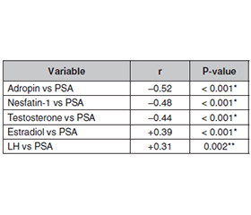
Актуальність. При раку передміхурової залози спостерігається метаболічний та гормональний дисбаланс, що впливає на розвиток пухлини. Регуляція гормонів відіграє життєво важливу роль у патофізіології раку передміхурової залози. Адропін, несфатин-1 і статеві гормони можуть бути використані як комбіновані біомаркери розвитку захворювання. Мета: оцінити інтегровану роль адропіну, несфатину-1 та статевих гормонів як метаболічних й ендокринних біомаркерів у розвитку і прогресуванні раку передміхурової залози. Матеріали та методи. Дослідження типу «випадок — контроль» було проведено з січня 2025 року до лютого 2026 року в іракських урологічних й онкологічних центрах за участю 100 пацієнтів із раком передміхурової залози та 50 здорових осіб контрольної групи. Діагноз було встановлено за допомогою визначення рівня простатспецифічного антигена (ПСА), ультразвукового обстеження й гістопатологічного аналізу (шкала Глісона). У дослідження були включені пацієнти віком 50 років і старше, у яких нещодавно діагностували рак, але вони не отримували лікування. Зразки крові (5 мл) центрифугували та зберігали при температурі –20 °C, а вміст адропіну і несфатину-1 визначали шляхом імуноферментного аналізу, тоді як статевих гормонів — за допомогою автоматизованого імуноферментного аналізатора. Клінічні дані були отримані з медичних записів та анкет. Результати. Не знайдено суттєвих відмінностей між показниками віку й статусу куріння. Однак індекс маси тіла, артеріальна гіпертензія та рівень ПСА були вірогідно підвищені в пацієнтів із раком. Біомаркери вірогідно відрізнялися (P = 0,001): рівень адропіну, несфатину-1 і тестостерону знижувався, а естрадіолу та лютеїнізуючого гормону — підвищувався. Ці показники були пов’язані з умістом ПСА і тяжкістю пухлинного процесу. Регресійний аналіз виявив низький рівень адропіну, несфатину-1, тестостерону, високі вміст естрадіолу й індекс маси тіла як незалежних предикторів ризику. Це вказує на роль метаболічно-ендокринної дисрегуляції в розвитку і прогресуванні раку передміхурової залози. Висновки. Метаболічний та ендокринний дисбаланс, який полягає у низькому рівні адропіну, несфатину-1 і тестостерону, а також підвищеному рівні естрадіолу, пов’язаний із раком передміхурової залози. Цей дисбаланс обумовлює процеси запалення, оксидативного стресу та порушення гормонального фону, що асоціюється з розвитком пухлини і погіршенням перебігу захворювання.
Background. In prostate cancer, there are metabolic and hormonal imbalances affecting the development of a tumor. Hormone regulation plays a vital role in the pathophysiology of prostate cancer. Adropin, nesfatin-1 and sex hormones can be used as combined biomarkers of disease development. The purpose of the study was to evaluate the integrated role of adropin, nesfatin-1, and sex hormones as metabolic-endocrine biomarkers in prostate cancer development and progression. Materials and methods. A case-control study was carried out between January 2025 and February 2026 in Iraqi urology and oncology centers involving 100 patients with prostate cancer and 50 healthy controls. They were diagnosed by means of prostate-specific antigen (PSA), ultrasound, and histopathology (Gleason score). Patients who were newly diagnosed and not treated, aged 50 years or older were eligible. Blood samples (5 mL) were centrifuged and stored at –20 °C, adropin and nesfatin-1 were evaluated by enzyme-linked immunosorbent assay whereas sex hormones were determined by an automated immunoassay system. Clinical data were obtained from records and questionnaires. Results. The findings indicated that there were no significant differences in age and smoking; hence, good matching. But body mass index, hypertension and PSA were all significantly elevated in patients. The biomarkers were all significantly different (P = 0.001) with decreased adropin, nesfatin-1 and testosterone and increased estradiol and luteinizing hormone. These indicators were associated with PSA and tumor severity. The regression analysis revealed low adropin, nesfatin-1, testosterone, high estradiol, and body mass index as the independent risk predictors that demonstrated the role of metabolic-endocrine dysregulation in the development and progression of prostate cancer. Conclusions. Metabolic-endocrine imbalance, which is low adropin, nesfatin-1, and testosterone, and elevated estradiol, are linked to prostate cancer. This imbalance facilitates the processes of inflammation, oxidative stress, and disruption of hormones, which are associated with tumor development and worsening of illnesses.
тестостерон; естрадіол; рак передміхурової залози; адропін; несфатин-1; метаболічні порушення
testosterone; estradiol; prostate cancer; adropin; nesfatin-1; metabolic disorders
Для ознакомления с полным содержанием статьи необходимо оформить подписку на журнал.
- Gandaglia G, et al. Epidemiology and prevention of prostate cancer. Eur Urol Oncol. 2021;4(6):877-892. doi: 10.1016/j.euo.2021.09.006.
- Berenguer CV, et al. Underlying features of prostate cancer-statistics, risk factors, and emerging methods for its diagnosis. Curr Oncol. 2023;30(2):2300-2321. doi: 10.3390/curroncol30020175.
- Adeloye D, et al. An estimate of the incidence of prostate cancer in Africa: a systematic review and meta-analysis. PLoS One. 2016;11(4):e0153496. doi: 10.1371/journal.pone.0153496.
- Marczuk N, et al. Adropin — physiological and pathophysiolo–gical role. Adv Hyg Exp Med (Postepy Hig Med Dosw). 2016;70:191-196. doi: 10.5604/17322693.1195845.
- Chen L, et al. Unveiling the multifaceted role of adropin in various diseases. Int J Mol Med. 2024;54(4):90. doi: 10.3892/ijmm.2024.5330.
- Niepolski L, Grzegorzewska AE. Salusins and adropin: new peptides potentially involved in lipid metabolism and atherosclerosis. Adv Med Sci. 2016;61(2):282-287. doi: 10.1016/j.advms.2016.03.004.
- Khalili S, et al. NUCB2/Nesfatin-1: a potent meal regulatory hormone and its role in diabetes. Egypt J Med Hum Genet. 2017;18(2):105-109. doi: 10.1016/j.ejmhg.2016.06.003.
- Dore R, et al. Nesfatin-1: functions and physiology of a novel regulatory peptide. J Endocrinol. 2017;232(1):R45-R65. doi: 10.1530/JOE-16-0361.
- Alemany M. The roles of androgens in humans: biology, meta–bolic regulation and health. Int J Mol Sci. 2022;23(19):11952. doi: 10.3390/ijms231911952.
- Crowley F, et al. A review of the pathophysiological mechanisms underlying castration-resistant prostate cancer. Res Rep Urol. 2021;13:457-472. doi: 10.2147/RRU.S269831.
- Rhee H, Vela I, Chung E. Metabolic syndrome and prostate cancer: a review of complex interplay amongst various endocrine factors in the pathophysiology and progression of prostate cancer. Horm Cancer. 2016;7(2):75-83. doi: 10.1007/s12672-015-0254-y.
- Di Sebastiano KM, et al. Glucose impairments and insulin resistance in prostate cancer: the role of obesity, nutrition and exercise. Obes Rev. 2018;19(7):1008-1016. doi: 10.1111/obr.12678.
- Gandhi J, et al. The molecular biology of prostate cancer: current understanding and clinical implications. Prostate Cancer Prostatic Dis. 2018;21(1):22-36. doi: 10.1038/s41391-017-0023-8.
- Perez-Cornago A, et al. Tall height and obesity are associated with an increased risk of aggressive prostate cancer: results from the EPIC cohort study. BMC Med. 2017;15(1):115. doi: 10.1186/s12916-017-0876-7.
- Zahid H, Simpson ER, Brown KA. Inflammation, dysregula–ted metabolism and aromatase in obesity and breast cancer. Curr Opin Pharmacol. 2016;31:90-96. doi: 10.1016/j.coph.2016.10.001.
- Divella R, et al. Obesity and cancer: the role of adipose tissue and adipocytokines-induced chronic inflammation. J Cancer. 2016;7(15):2346. doi: 10.7150/jca.16884.
- Liang Z, et al. Hypertension and risk of prostate cancer: a systematic review and meta-analysis. Sci Rep. 2016;6:31358. doi: 10.1038/srep31358.
- Petrelli F, et al. Effects of hypertension on cancer survival: a meta-analysis. Eur J Clin Invest. 2021;51(6):e13493. doi: 10.1111/eci.13493.
- Claps M, et al. Testosterone levels and prostate cancer prognosis: systematic review and meta-analysis. Clin Genitourin Cancer. 2018;16(3):165-175. doi: 10.1016/j.clgc.2017.12.012.
- Hauger RL, et al. The role of testosterone, the androgen receptor, and hypothalamic-pituitary-gonadal axis in depression in ageing men. Rev Endocr Metab Disord. 2022;23(6):1259-1273. doi: 10.1007/s11154-022-09720-2.
- Kumar R, et al. The testosterone paradox of advanced prostate cancer: mechanistic insights and clinical implications. Nat Rev Urol. 2023;20(5):265-278. doi: 10.1038/s41585-023-00674-3.
- Chinapayan SM, et al. Potential value of visfatin, omentin-1, nesfatin-1 and apelin in renal cell carcinoma: a systematic review and meta-analysis. Diagnostics. 2022;12(12):3069. doi: 10.3390/diagnostics12123069.
- Pawlowska-Olszewska M, et al. Adropin, nesfatin-1 and angiotensin II receptor expression in the abdominal aorta in ovariectomized rats after nesfatin-1 treatment. J Physiol Pharmacol. 2019;70(6). doi: 10.26402/jpp.2019.6.05.
- Ragab A, et al. Serum nesfatin-1 level in men with diabetes and erectile dysfunction correlates with generalized anxiety disorder-7: a prospective comparative study. Andrology. 2023;11(2):307-315. doi: 10.1111/andr.13345.
- Weng WC, et al. Potential impact of omentin-1 genetic variants on perineural invasion in prostate cancer. J Cancer. 2025;16(12):3767. doi: 10.7150/jca.
- Schneider G, et al. Tissue-specific tumorigenesis: context matters. Nat Rev Cancer. 2017;17(4):239-253. doi: 10.1038/nrc.2017.5.
- Wang Y, et al. Expression of serum PSA, nesfatin-1, and AMH in patients with polycystic ovary syndrome. Cell Mol Biol. 2021;67(5):57-63. doi: 10.14715/cmb/2021.67.5.8.
- Wang M, Tong J, Zhu Q, Tang H, Tang L. Blood nesfatin-1 levels in patients with polycystic ovary syndrome: a systematic review and meta-analysis. Front Endocrinol (Lausanne). 2024 Jan 24;14:1275753. doi: 10.3389/fendo.2023.1275753.
- Mohan H, Ramesh N, Mortazavi S, Le A, Iwakura H, Unniappan S. Nutrients differentially regulate nucleobindin-2/nesfatin-1 in vitro in cultured stomach ghrelinoma (MGN3-1) cells and in vivo in male mice. PLoS One. 2014 Dec 15;9(12):e115102. doi: 10.1371/journal.pone.0115102.
- Koç A, et al. Association between serum NUCB2/nesfatin-1 levels and erectile dysfunction. Exp Ther Med. 2024;28(5):428. doi: 10.3892/etm.2024.
- Pascuzzi N, et al. Recent breakthroughs in breast cancer endocrinology and tumor microenvironmental interactions. In: Latest Research on Breast Cancer — Molecular Insights, Diagnostic Advan–ces and Therapeutic Innovations. IntechOpen; 2024. doi: 10.5772/intechopen.