
Журнал «Медицина неотложных состояний» Том 22, №5, 2026
Вернуться к номеру
Тепловізійна оцінка тяжкості термічної травми на етапах спеціалізованого лікування
Авторы: Олійник Г.А. (1), Шустакова Г.В. (2), Фоменко Ю.В. (2), Гордієнко Е.Ю. (2), Кравцов О.В. (3, 4), Грязін О.Є. (1)
(1) - Харківський національний медичний університет, м. Харків, Україна
(2) - Фізико-технічний інститут низьких температур імені Б.І. Вєркіна НАН України, м. Харків, Україна
(3) - ДУ «Інститут загальної та невідкладної хірургії імені В.Т. Зайцева НАМН України», м. Харків, Україна
(4) - КНП «Міська клінічна лікарня швидкої та невідкладної медичної допомоги імені проф. О.І. Мещанінова», м. Харків, Україна
Рубрики: Медицина неотложных состояний
Разделы: Клинические исследования
Версия для печати
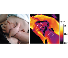
Актуальність. Інфрачервона термографія визнається корисним допоміжним методом оцінки опікових ран, хоча кількісна інтерпретація термограм залишається недостатньо стандартизованою та потребує вдосконалення. Мета дослідження: оптимізувати обробку термограм, встановити температурні критерії для диференціації ділянок опіків різної глибини та розробити практичну методику термографічної оцінки та моніторингу опікових ран. Матеріали та методи. Дослідження проводилося в опіковому відділенні Харківської міської клінічної лікарні невідкладної та екстреної медичної допомоги імені професора О.І. Мещанінова протягом 2021–2025 років. Було періодично термографовано 192 пацієнтів з опіками різної етіології, локалізації, глибини та ступеня. Дані 121 пацієнта (295 опікових ран), обстежених у перші три доби після опіку, використано для розробки діагностичного тесту та визначення температур відсікання глибоких, межових та поверхневих опіків. Використовувалися непараметричні статистичні методи та ROC-аналіз. Результати. Термічні параметри, отримані з опіків різної глибини, продемонстрували статистично значущі відмінності та дозволили розробити двопороговий класифікаційний підхід. ROC-аналіз визначив порогове значення температури ΔTcut-off = –2,3 °C для диференціації глибоких опіків від інших травм, з чутливістю 83,3 %, специфічністю 88,9 %, AUC = 0,954. Порогове значення ΔTcut-off = 0 °C диференціювало поверхневі опіки від інших травм з чутливістю 72,7 %, специфічністю 89,5 % та AUC = 0,931. На основі результатів діагностичного тесту було розроблено методику оптимізованої обробки термограм, яка дозволяє підвищити точність оцінки опікової рани. Висновки. Запропонована методика поліпшує кількісну інтерпретацію термограм та підвищує точність оцінки опікових ран. Її застосування може сприяти плануванню лікувальної тактики та здійсненню термографічного контролю перебігу ранового процесу на різних етапах надання медичної допомоги.
Background. Infrared thermography is recognised as a useful modality for burn wound assessment, although quantitative interpretation of thermograms remains insufficiently standardised and requires refinement. The aim of the study was to optimise thermogram processing, establish temperature criteria for differentiating burn areas with different depths, and to develop a practical procedure for thermographic assessment and monitoring of burn wounds. Materials and methods. The study was carried out in the burn unit of the Kharkiv City Clinical Hospital for Emergency Medical Services named after prof. O.I. Meschaninov during 2021–2025. A total of 192 patients with burns of various origins, locations, depths and extent were examined by thermography throughout hospitalisation. Data from 121 patients (295 burn wounds) examined within the first three days after injury were used to develop a diagnostic test and determine cut-off temperatures for deep, borderline and superficial burns. Nonparametric statistical methods and ROC analysis were used. Results. Thermal parameters obtained from burns of different depths demonstrated statistically significant differences and enabled development of a two-threshold classification approach. ROC analysis identified a threshold temperature value of ΔTcut-off = –2.3 °C for differentiating deep burns from other injuries, with a sensitivity of 83.3 %, specificity of 88.9 %, AUC = 0.954. A threshold value of ΔTcut-off = 0 °C differentiated superficial burns from other injuries, with a sensitivity of 72.7 %, specificity of 89.5 %, and AUC = 0.931. Based on the results of diagnostic test, a method for optimized thermogram processing was developed that allows us to increase the accuracy of burn wound assessment. Conclusions. The proposed procedure improves quantitative interpretation of thermograms and increases the accuracy of burn wound assessment. It may facilitate treatment planning and thermographic control of the wound process at different stages of care.
термічна травма; глибина опіку; інфрачервона термографія
thermal trauma; burn depth; infrared thermography
Для ознакомления с полным содержанием статьи необходимо оформить подписку на журнал.
- Merchant M, Hu SB, Miller C, Ahmadi T, Garcia E, Smith MI. Comprehensive Management of Severe Burn Injuries: A Multidisciplinary Approach from Resuscitation to Rehabilitation. Emerg Care Med. 2025;2:26-46. https://doi.org/10.3390/ecm2020026.
- Markiewicz-Gospodarek A, Kozioł M, Tobiasz M, Baj J, Radzikowska-Büchner E, Przekora A. Burn Wound Healing: Clinical Complications, Medical Care, Treatment, and Dressing Types: The Current State of Knowledge for Clinical Practice. Int J Environ Res Public Health. 2022;19(3):1338-63. https://doi.org/10.3390/ijerph19031338.
- Dobson GP, Morris JL, Letson HL. Pathophysiology of Severe Burn Injuries: New Therapeutic Opportunities From a Systems Perspective. J Burn Care Res. 2024;45(4):1041-50. https://doi.org/10.1093/jbcr/irae049.
- Jeschke MG, van Baar ME, Choudhry MA, Chung KK, Gibran NS, Logsetty S. Burn injury. Nat Rev Dis Primers. 2020;6:11. https://doi.org/10.1038/s41572-020-0145-5.
- Sarda NN, Hingway S. Screening and Phasewise Management of Burn Injuries. Cureus. 2024;16(2):e54915. doi: 10.7759/cureus.54915.
- Walker NJ, King KC. Acute and Chronic Thermal Burn Evaluation and Management. In: StatPearls [Internet]. Treasure Island (FL): StatPearls Publishing; 2026. Available from: https://www.ncbi.nlm.nih.gov/books/NBK430730/. Accessed: June19, 2026.
- Jaspers MEH, van Haasterecht L, van Zuijlen PPM, Mokkink LB. A systematic review on the quality of measurement techniques for the assessment of burn wound depth or healing potential. Burns. 2019;45(2):261-81. https://doi.org/10.1016/j.burns.2018.05.015.
- Monstrey S, Hoeksema H, Verbelen J, Pirayesh A, Blondeel P. Assessment of burn depth and burn wound healing potential. Burns. 2008;34(6):761-9. https://doi.org/10.1016/j.burns.2008.01.009.
- Carrière ME, de Haas LEM, Pijpe A, Meij-de Vries A, Gardien KLM, van Zuijlen PPM, et al. Validity of thermography for measuring burn wound healing potential. Wound Repair Regen. 2020;28(3):347-54. https://doi.org/10.1111/wrr.12786.
- Dang J, Lin M, Tan C, Pham CH, Huang S, Hulsebos IF, Yenikomshian H, Gillenwater J. Use of Infrared Thermography for Assessment of Burn Depth and Healing Potential: A Systematic Review. J Burn Care Res. 2021;42(6):1120-7. https://doi.org/10.1093/jbcr/irab108.
- Zheng KJ, Middelkoop E, Stoop M, van Zuijlen PPM, Pijpe A. Validity of laser speckle contrast imaging for the prediction of burn wound healing potential. Burns. 2022;48(2):319-27. https://doi.org/10.1016/j.burns.2021.04.028.
- Wearn C, Lee KC, Hardwicke J, Allouni A, Bamford A, Nightingale P, et al. Prospective comparative evaluation study of Laser Doppler Imaging and thermal imaging in the assessment of burn depth. Burns. 2018;44(1):124-33. https://doi.org/10.1016/j.burns.2017.08.004.
- Asif A, Poyiatzis C, Raheman FJ and Rojoa DM. The Use of Infrared Thermography (IRT) in Burns Depth Assessment: A Diagnostic Accuracy Meta-Analysis. Eur Burn J. 2022;3(3):432-46. https://doi.org/10.3390/ebj3030038.
- Jaspers MEH, Maltha I, Klaessens JHGM, de Vet HCW, Verdaasdonk RM, van Zuijlen PPM. Insights into the use of thermography to assess burn wound healing potential: a reliable and valid technique when compared to laser Doppler imaging. J Biomed Opt. 2016;21(9):e096006. doi: 10.1117/1.JBO.21.9.096006.
- Martınez-Jimenez MA, Ramirez-GarciaLuna JL, Kolosovas-Machuca ES, Drager J, Gonzalez FJ. Development and validation of an algorithm to predict the treatment modality of burn wounds using thermographic scans: Prospective cohort study. PLoS One. 2018;13(11):e0206477. https://doi.org/10.1371/journal.pone.0206477.
- Medina-Preciado JD, Kolosovas-Machuca ES, Velez-Gomez E, Miranda-Altamirano A, González FJ. Noninvasive determination of burn depth in children by digital infrared thermal ima–ging. J Biomed Opt. 2013;18(6):e061204. https://doi.org/10.1117/1.JBO.18.6.061204.
- Miccio J, Parikh S, Marinaro X, Prasad A, McClain S, Singer AJ, Clark RA. Forward-looking infrared imaging predicts ultimate burn depth in a porcine vertical injury progression model. Burns 2016;42(2):397-404. https://doi.org/10.1016/j.burns.2015.07.006.
- Singer AJ, Relan P, Beto L, Jones-Koliski L, Sandoval S, Clark RAF. Infrared Thermal Imaging Has the Potential to Reduce Unnecessary Surgery and Delays to Necessary Surgery in Burn Patients. J Burn Care Res. 2016:37(6):350-5. https://doi.org/10.1097/BCR.0000000000000330.
- Ammer K. Glamorgan Protocol for recording and evaluation of thermal images of the human body. Thermol Int. 2008;18(4):125-9. Available from: https://www.researchgate.net/publication/233420893_The_Glamorgan_Protocol_for_recording_and_evaluation_of_thermal_images_of_the_human_body. Accessed: June19, 2026.
- Becerra AG, Tiznado JEO, Alcaraz JLG, Wilson CC, Barreras JAL, Gutiérrez JCC, et al. Temperature Asymmetry Analysis between Left and Right Wrist with Sensory and Infrared Thermo–graphy. Int J Environ Res. Public Health. 2022;19:10240. https://doi.org/10.3390/ijerph191610240.
- Vardasca R, Ring F, Plassmann P, Jones C. Thermal symmetry of the upper and lower extremities in healthy subjects. Thermol Int. 12;22(2):53-60. Available from: https://www.researchgate.net/publication/227860586. Accessed June19, 2026.
- Burke-Smith A, Collier J, Jones I. A comparison of non-invasive imaging modalities: Infrared thermography, spectrophotometric intracutaneous analysis and laser Doppler imaging for the assessment of adult burns. Burns. 2015;41(8):1695-707. https://doi.org/10.1016/j.burns.2015.06.023.
- Hardwicke J, Thomson R, Bamford A, Moiemen N. A pilot evaluation study of high resolution digital thermal imaging in the assessment of burn depth. Burns. 2013;39(1):76-81. https://doi.org/10.1016/j.burns.2012.03.014.
- Jaspers MEH, Carrière ME, Meij-de Vries A, Klaessens JHGM, van Zuijlen PPM. The FLIR ONE thermal imager for the assessment of burn wounds: Reliability and validity study. Burns. 2017;43(7):1516-23. https://doi.org/10.1016/j.burns.2017.04.006.
- Hargroder AG, Davidson JE Sr, Luther DG, Head JF. Infrared imaging of burn wounds to determine burn depth. Proc. SPIE 3698, Infrared Technology and Applications XXV (26 July 1999). https://doi.org/10.1117/12.354509.
- Ganon S, Guédon A, Cassier S, Atlan M. Contribution of thermal imaging in determining the depth of pediatric acute burns. Burns. 2020;46:1091-9. https://doi.org/10.1016/j.burns.2019.11.019.
- Hackett MEJ. The use of thermography in the assessment of burn and blood supply of flaps, with preliminary reports on its use in Dupuytren’s contracture and treatment of varicose ulcers. Br J Plast Surg. 1974;27:311-7. https://doi.org/10.1016/0007-1226(74)90028-9.
- Madian IM, Sherif WI, El Fahar MH, Othman WN. The use of smartphone thermography to evaluate wound healing in second-degree burns. Burns. 2025;51(3):107307. https://doi.org/10.1016/j.burns.2024.107307.
- Glushchuk M, Shustakova G, Gordiyenko E, Fomenko Y, Oliynyk G, Dvortcevyi V, et al. Thermal Imaging Study of Human Soft Tissue Lesions and Biological Tissue Exposure to Low-Temperature in vivo. Sci. Innov. 2022;19(6):83-96. https://doi.org/10.15407/scine18.06.083.
- Badea M-S, Vertan C, Florea C, Florea L, Bădoiu S. Severe burns assessment by joint color-thermal imagery and ensemble me–thods. In: 2016 IEEE 18th International Conference on e-Health Networking, Applications and Services (HealthCom). Munich, Germany; 2016. Р. 1-5. doi: 10.1109/HealthCom.2016.7749450.