
Международный эндокринологический журнал Том 22, №6, 2026
Вернуться к номеру
Рівень інтерлейкіну-10 та -22 у крові і тканинах пацієнтів із медулярною та папілярною карциномами щитоподібної залози
Авторы: P.P. Zinych, M.Yu. Bolhov
V.P. Komisarenko Institute of Endocrinology and Metabolism of NAMSU, Kyiv, Ukraine
Рубрики: Эндокринология
Разделы: Клинические исследования
Версия для печати
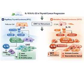
Актуальність. Взаємодія пухлини та імунної системи критично впливає на прогресування раку щитоподібної залози (ЩЗ). Серед імунорегуляторних цитокінів інтерлейкін-10 (IL-10) та IL-22 виконують парадоксальні функції, пригнічуючи протипухлинний імунітет, одночасно сприяючи виживанню й метастазуванню пухлинних клітин. Однак їх тканинний розподіл і роль у метастазуванні залишаються недостатньо з’ясованими. Мета: провести порівняльну оцінку вмісту IL-10 та IL-22 в умовно нормальній (УН) тканині ЩЗ, тканинах первинної медулярної (МКЩЗ) та папілярної карциноми ЩЗ (ПКЩЗ), а також у плазмі периферичної крові пацієнтів із метастатичним і неметастатичним перебігом захворювання. Матеріали та методи. Зразки пухлини ЩЗ, УН тканини, метастатичних уражень та плазми периферичної крові були отримані від пацієнтів після хірургічного лікування. Концентрації IL-10 та IL-22 визначали кількісно за допомогою імуноферментного аналізу. Статистичні порівняння проводили за допомогою параметричних і непараметричних тестів. Результати. Концентрації IL-10 були вірогідно підвищені в тканинах МКЩЗ та метастазів порівняно з відповідними УН зразками. У пацієнтів із метастатичною ПКЩЗ уміст IL-10 як у тканинах, так і у плазмі був вірогідно вищий, ніж в осіб із ПКЩЗ без метастазів. Концентрації IL-22 у тканинах неметастатичної МКЩЗ перевищували такі в прилеглій УН тканині майже у 2 рази. Найвищий рівень IL-22 виявлено в метастатичних ураженнях лімфатичних вузлів при МКЩЗ, що свідчить про поступове його зростання вздовж осі «первинна пухлина — метастази». Висновки. Таким чином, показник IL-10 у плазмі пацієнтів із ПКЩЗ може бути передопераційним маркером утворення метастазів. Рівень IL-22 у післяопераційній тканині хворих із ПКЩЗ може бути маркером появи метастазів у майбутньому, а його вміст у плазмі пацієнтів із МКЩЗ можна використовувати як передопераційний предиктор утворення метастазів. Це вказує на те, що IL-22 є потенційним біомаркером для стратифікації ризику метастазування при карциномах ЩЗ.
Background. Tumor-immune interactions critically shape thyroid cancer progression. Among immunoregulatory cytokines, interleukin-10 (IL-10) and IL-22 exert paradoxical functions by suppressing anti-tumor immunity while promoting tumor cell survival and metastasis (Mts). Yet the tissue-systemic distribution and metastatic relevance of IL-10 and IL-22 remain insufficiently defined. The aim of the study was to comparatively evaluate IL-10 and IL-22 levels in conditionally normal (CN) thyroid tissue, primary medullary thyroid carcinoma (MTC) and papillary thyroid carcinoma (PTC) tissues, as well as in peripheral blood plasma of patients with metastatic and non-metastatic disease. Materials and methods. Postoperative tumor, CN thyroid tissue, metastatic lesions, and plasma samples were obtained from surgically treated patients. IL-10 and IL-22 concentrations were quantified by enzyme-linked immunosorbent assay. Statistical comparisons were performed using parametric and nonparametric tests. Results. IL-10 levels were significantly elevated in MTC and Mts tissues compared to corresponding CN thyroid samples. In patients with metastatic PTC, IL-10 concentrations both in tissues and plasma were significantly higher than in non-metastatic PTC samples. IL-22 concentrations in the non-metastatic MTC exceeded those of adjacent CN tissue by almost 2-fold. The highest IL-22 concentrations were detected in metastatic lymph node lesions of MTC, indicating progressive upregulation along the primary tumor-Mts axis. Conclusions. Thus, level of IL-10 in plasma of patients with PTC can be preoperative marker predicting Mts formation. The level of IL-22 in the postoperative tissue of patients with PTC can be marker predicting Mts formation in a future, and the level of IL-22 in plasma of patients with MTC can be used as preoperative marker predicting Mts formation. These findings implicate as a potential biomarker for stratifying metastatic risk in thyroid carcinomas.
інтерлейкін-10; інтерлейкін-22; медулярна та папілярна карциноми щитоподібної залози; метастазування; біомаркери
interleukin-10; interleukin-22; medullary and papillary thyroid carcinomas; metastasis; biomarkers
Для ознакомления с полным содержанием статьи необходимо оформить подписку на журнал.
- Saraiva M, Vieira P, O’Garra A. Biology and therapeutic potential of interleukin-10. J Exp Med. 2020 Jan 6;217(1):e20190418. doi: 10.1084/jem.20190418.
- Chang CM, Lam HYP, Hsu HJ, Jiang SJ. Interleukin-10: A double-edged sword in breast cancer. Tzu Chi Med J. 2021 Feb 24;33(3):203-211. doi: 10.4103/tcmj.tcmj_162_20.
- Loonen AJM. Putative role of immune reactions in the mecha–nism of tardive dyskinesia. Brain Behav Immun Health. 2023 Sep 23;33:100687. doi: 10.1016/j.bbih.2023.100687.
- Shiri AM, Fard-Aghaie M, Bedke T, et al. Foxp3 + Treg-derived IL-10 promotes colorectal cancer-derived lung metastasis. Sci Rep. 2024 Dec 16;14(1):30483. doi: 10.1038/s41598-024-80437-8.
- Shiri AM, Zhang T, Bedke T, et al. IL-10 dampens antitumor immunity and promotes liver metastasis via PD-L1 induction. J Hepatol. 2024 Apr;80(4):634-644. doi: 10.1016/j.jhep.2023.12.015.
- Guo Z, He Q, Zhang Y, Li Y, Zhang Z. Mechanisms of Interleukin-10-Mediated Immunosuppression in Viral Infections. Pathogens. 2025 Oct 1;14(10):989. doi: 10.3390/pathogens14100989.
- Ouyang W, O’Garra A. IL-10 Family Cytokines IL-10 and IL-22: from Basic Science to Clinical Translation. Immunity. 2019 Apr 16;50(4):871-891. doi: 10.1016/j.immuni.2019.03.020.
- Wei HX, Wang B, Li B. IL-10 and IL-22 in Mucosal Immunity: Driving Protection and Pathology. Front Immunol. 2020 Jun 26;11:1315. doi: 10.3389/fimmu.2020.01315.
- Li J, Wu Z, Wu Y, et al. IL-22, a vital cytokine in autoimmune diseases. Clin Exp Immunol. 2024 Nov 12;218(3):242-263. doi: 10.1093/cei/uxae035.
- Zheng Y, Li T. Interleukin-22, a potent target for treatment of non-autoimmune diseases. Hum Vaccin Immunother. 2018;14(12):2811-2819. doi: 10.1080/21645515.2018.1509649.
- Dudakov JA, Hanash AM, van den Brink MR. Interleukin-22: immunobiology and pathology. Annu Rev Immunol. 2015;33:747-85. doi: 10.1146/annurev-immunol-032414-112123.
- Hunzeker ZE, Zhao L, Kim AM, et al. The role of IL-22 in cancer. Med Oncol. 2024 Sep 5;41(10):240. doi: 10.1007/s12032-024-02481-8.
- Briukhovetska D, Suarez-Gosalvez J, Voigt C, et al. T cell-derived interleukin-22 drives the expression of CD155 by cancer cells to suppress NK cell function and promote metastasis. Immunity. 2023 Jan 10;56(1):143-161.e11. doi: 10.1016/j.immuni.2022.12.010.
- Bradford MM. A rapid and sensitive method for the quantitation of microgram quantities of protein utilizing the principle of protein-dye binding. Anal Biochem. 1976 May 7;72:248-254. doi: 10.1016/0003-2697(76)90527-3.
- Ni G, Zhang L, Yang X, et al. Targeting interleukin-10 signalling for cancer immunotherapy, a promising and complicated task. Hum Vaccin Immunother. 2020 Oct 2;16(10):2328-2332. doi: 10.1080/21645515.2020.1717185.
- Saxton RA, Tsutsumi N, Su LL, et al. Structure-based decoupling of the pro- and anti-inflammatory functions of interleukin-10. Science. 2021 Mar 19;371(6535):eabc8433. doi: 10.1126/science.abc8433.
- Carlini V, Noonan DM, Abdalalem E, et al. The multifaceted nature of IL-10: regulation, role in immunological homeostasis and its relevance to cancer, COVID-19 and post-COVID conditions. Front Immunol. 2023 Jun 8;14:1161067. doi: 10.3389/fimmu.2023.1161067.
- Salkeni MA, Naing A. Interleukin-10 in cancer immunothe–rapy: from bench to bedside. Trends Cancer. 2023 Sep;9(9):716-725. doi: 10.1016/j.trecan.2023.05.003.
- El-Shemi AG, Alqurashi A, Abdulrahman JA, et al. IL-10-Directed Cancer Immunotherapy: Preclinical Advances, Clini–cal Insights, and Future Perspectives. Cancers (Basel). 2025 Mar 17;17(6):1012. doi: 10.3390/cancers17061012.
- Zeng C, Niu F, Li H, et al. Progress of IL-10 and li–ver metastasis. Cytokine. 2025 Jun;190:156932. doi: 10.1016/j.cyto.2025.156932.
- Wang Y, Liu XH, Li YH, Li O. The paradox of IL-10-mediated modulation in cervical cancer (Review). Biomed Rep. 2013;1:347-351. doi: 10.3892/br.2013.69.
- Mannino MH, Zhu Z, Xiao H, Bai Q, Wakefield MR, Fang Y. The paradoxical role of IL-10 in immunity and cancer. Cancer Lett. 2015 Oct 28;367(2):103-7. doi: 10.1016/j.canlet.2015.07.009.
- Li B, Wang F, Ma C, Hao T, Geng L, Jiang H. Predictive va–lue of IL-18 and IL-10 in the prognosis of patients with colorectal cancer. Oncol Lett. 2019 Jul;18(1):713-719. doi: 10.3892/ol.2019.10338.
- Aslam A, Refaat B, Almaimani RA, et al. Increased protein expression of interleukin-10 and its signalling molecules in colon cancer progression: potential prognostic and therapeutic targets. Discov Oncol. 2025 Apr 29;16(1):637. doi: 10.1007/s12672-025-02452-z.
- Yu J, Feng L, Luo Z, et al. Interleukin-10 deficiency suppresses colorectal cancer metastasis by enriching gut Parabacteroides distasonis. J Adv Res. 2025 Oct;76:467-479. doi: 10.1016/j.jare.2024.11.024.
- Sullivan KM, Jiang X, Guha P, et al. Blockade of interleukin 10 potentiates antitumour immune function in human colorectal cancer liver metastases. Gut. 2023 Feb;72(2):325-337. doi: 10.1136/gutjnl-2021-325808.
- Cunha LL, Morari EC, Nonogaki S, et al. Interleukin 10 expression is related to aggressiveness and poor prognosis of patients with thyroid cancer. Cancer Immunol Immunother. 2017;66:141-8. doi: 10.1007/s00262-016-1924-4.
- Xi C, Zhang GQ, Sun ZK, et al. Interleukins in Thyroid Cancer: From Basic Researches to Applications in Clinical Practice. Front Immunol. 2020 Jun 12;11:1124. doi: 10.3389/fimmu.2020.01124.
- Menicali E, Guzzetti M, Morelli S, Moretti S, Puxeddu E. Immune Landscape of Thyroid Cancers: New Insights. Front Endocrinol (Lausanne). 2021 Apr 27;11:637826. doi: 10.3389/fendo.2020.637826.
- Stassi G, Todaro M, Zerilli M, et al. Thyroid cancer resistance to chemotherapeutic drugs via autocrine production of interleukin-4 and interleukin-10. Cancer Res. 2003;63(20):6784-6790. doi: 10.1016/S0165-4608(02)00795-1.
- Todaro M, Zerilli M, Ricci-Vitiani L, et al. Autocrine production of interleukin-4 and interleukin-10 is required for survival and growth of thyroid cancer cells. Cancer Res. 2006;66(3):1491-1499. doi: 10.1158/0008-5472.CAN-05-2514.
- Provatopoulou X, Georgiadou D, Sergentanis TN, et al. Interleukins as markers of inflammation in malignant and benign thyroid disease. Inflamm Res. 2014 Aug;63(8):667-74. doi: 10.1007/s00011-014-0739-z.
- Martins MB, Marcello MA, Batista FA, et al. Serum interleukin measurement may help identify thyroid cancer patients with active disease. Clin Biochem. 2018 Feb;52:1-7. doi: 10.1016/j.clinbiochem.2017.10.003.
- Denaro N, Romanò R, Alfieri S, et al. The Tumor Microenvironment and the Estrogen Loop in Thyroid Cancer. Cancers (Basel). 2023 Apr 25;15(9):2458. doi: 10.3390/cancers15092458.
- Çil E, Kumral A, Kanmaz-Özer M, et al. Interleukin-10-1082 gene polymorphism is associated with papillary thyroid cancer. Mol Biol Rep. 2014 May;41(5):3091-7. doi: 10.1007/s11033-014-3169-7.
- Yu Y, Qiao M, Liu J, Guo Y, Sun Y. The reciprocal regulation between autophagy and IL-22: implications for immunity and therapy. Clin Exp Med. 2025 Jun 4;25(1):187. doi: 10.1007/s10238-025-01695-y.
- Kirchberger S, Royston DJ, Boulard O, et al. Innate lymphoid cells sustain colon cancer through production of interleukin-22 in a mouse model. J Exp Med. 2013 May 6;210(5):917-31. doi: 10.1084/jem.20122308.
- Giannou AD, Kempski J, Shiri AM, et al. Tissue resident iNKT17 cells facilitate cancer cell extravasation in liver metastasis via interleukin-22. Immunity. 2023 Jan 10;56(1):125-142.e12. doi: 10.1016/j.immuni.2022.12.014.
- Markota A, Endres S, Kobold S. Targeting interleukin-22 for cancer therapy. Hum Vaccin Immunother. 2018;14(8):2012-2015. doi: 10.1080/21645515.2018.1461300.
- Mei Z, Zhou L, Zhu Y, et al. Interleukin-22 promotes pa–pillary thyroid cancer cell migration and invasion through micro–RNA-595/Sox17 axis. Tumour Biol. 2016;37:11753-62. doi: 10.1007/s13277-016-5030-1.
- Qing C, Ghorani E. Two faces: IL-22 effects prevail over defense against metastasis. Immunity. 2023 Jan 10;56(1):6-8. doi: 10.1016/j.immuni.2022.12.013.
- Lim C, Savan R. The role of the IL-22/IL-22R1 axis in cancer. Cytokine Growth Factor Rev. 2014 Jun;25(3):257-71. doi: 10.1016/j.cytogfr.2014.04.005.
- Kryczek I, Lin Y, Nagarsheth N, et al. IL-22+CD4+ T cells promote colorectal cancer stemness via STAT3 transcription factor activation and induction of the methyltransferase DOT1L. Immunity. 2014 May 15;40(5):772-784. doi: 10.1016/j.immuni.2014.03.010.
- Cui X, Jing X, Yi Q, et al. IL22 furthers malignant transformation of rat mesenchymal stem cells, possibly in association with IL22RA1/STAT3 signaling. Oncol Rep. 2019 Apr;41(4):2148-2158. doi: 10.3892/or.2019.7007.
- Zinych PP, Pushkarev VM, Levchuk NI, Tronko MD. ZEB1 as a marker of metastasis in thyroid carcinomas: review of literature and own data. Mìžnarodnij endokrinologìčnij žurnal. 2024;20(7):517-528. doi: 10.22141/2224-0721.20.7.2024.1451 (in Ukrainian).
- Li W, Li F, Lei W, Tao Z. TRIM30 modulates Interleukin-22-regulated papillary thyroid Cancer cell migration and invasion by targeting Sox17 for K48-linked Polyubiquitination. Cell Commun Signal. 2019 Dec 10;17(1):162. doi: 10.1186/s12964-019-0484-6.
- Zhang T, Wahib R, Zazara DE, et al. CD4+ T cell-derived IL-22 enhances liver metastasis by promoting angiogenesis. Oncoimmunology. 2023 Oct 20;12(1):2269634. doi: 10.1080/2162402X.2023.2269634.
- Ruan HX, Fang YL, Qin XN, Lin L. Interleukin-22 promotes cancer stemness and chemotherapy resistance in colorectal cancer via epidermal growth factor receptor/extracellular signal-regulated kinase pathway. World J Gastrointest Oncol. 2025 Aug 15;17(8):108535. doi: 10.4251/wjgo.v17.i8.108535.