
Международный эндокринологический журнал Том 22, №6, 2026
Вернуться к номеру
Оцінка впливу аполіпопротеїну J і CX3CL1 на розвиток і прогресування діабетичної ретинопатії на основі математичного моделювання
Авторы: Венгер А.Ю.
Одеський національний медичний університет, м. Одеса, Україна
Рубрики: Эндокринология
Разделы: Клинические исследования
Версия для печати
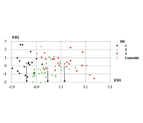
Актуальність. Результати клінічних та експериментальних досліджень свідчать про участь хемокінів аполіпопротеїну J і CX3CL1 у патогенезі діабетичних уражень сітківки. Можливий лікувальний потенціал аполіпопротеїну J і CX3CL1 спонукає до подальших поглиблених клінічних досліджень їхньої прогностичної значущості, особливо на основі математичного моделювання. Мета роботи полягала в розробці математичної моделі оцінки прогностичної значущості аполіпопротеїну J і CX3CL1 у розвитку та прогресуванні діабетичної ретинопатії (ДР) на основі дискримінантного аналізу. Матеріали та методи. Дослідження було відкритим, одномоментним, когортним, неінтервенційним, за типом «випадок — контроль», одноцентровим. Критерії включення пацієнтів у дослідження: громадяни України (українська популяція), інформована згода пацієнта, документальне підтвердження діагнозу цукрового діабету (ЦД) 2-го типу, вік від 18 років, дотримання положень Гельсінської декларації Всесвітньої медичної асоціації (Сеул, 2008). Офтальмологічні критерії невключення: патологія зорового нерва, макулопатія, глаукома, зріла катаракта, гемофтальм, відшарування сітківки різного генезу. Для розробки математичної моделі для аполіпопротеїну J і CX3CL1 було класифіковано 18 хворих на ЦД 2-го типу і непроліферативну ДР (І стадія, група 1), 42 хворих на ЦД 2-го типу і препроліферативну ДР (ІІ стадія, група 2) і 22 хворих на ЦД 2-го типу і проліферативну ДР (ІІІ стадія, група 3). Головним статистичним методом обробки результатів був дискримінантний аналіз. Значення P < 0,05 вважалося статистично значущим. Для статистичної обробки отриманих даних застосовувався програмний пакет Statistica 9.0 (StatSoft, Tulsa, OK, USA). Результати. Проведений аналіз дозволив розробити математичний алгоритм для оцінки ризику обтяження ДР у хворих на ЦД 2-го типу за участі аполіпопротеїну J і CX3CL1. В основі складання прогнозу перебігу ДР лежить обчислення функцій класифікації (ФК), які були отримані за результатами побудови математичної моделі. За отриманими клінічними даними й лабораторними показниками підраховуються ФК1, ФК2 і ФК3. Далі порівнюють ФК1 з ФК2 або ФК2 з ФК3. Прогностичне рішення приймається як вибір тієї ФК, яка має більше значення. Так, якщо ФК1 > ФК2, то прогноз позитивний (стабілізація процесу на І або ІІ стадії ДР). Якщо ФК2 > ФК3, то прогноз відносно сприятливий (стабілізація процесу на ІІ стадії ДР). Якщо ФК1 < ФК2 або ФК2 < ФК3, то прогноз негативний, є імовірність ризику обтяження ДР і прогресування ДР на наступну стадію. Висновки. Модель оцінки прогностичної значущості аполіпопротеїну J має інформативність 74,39 % і вірогідність Р = 0,0001. Модель оцінки прогностичної значущості CX3CL1 має інформативність 78,5 % і вірогідність Р = 0,00001.
Background. The results of clinical and experimental studies indicate the involvement of chemokines apolipoprotein J and CX3CL1 in the pathogenesis of diabetic retinopathy (DR). The potential therapeutic relevance of these molecules warrants further in-depth clinical investigations aimed at evaluating their prognostic significance, especially based on mathematical modeling. The purpose of the work was to develop a mathematical model for assessing the prognostic significance of apolipoprotein J and CX3CL1 in the development and progression of DR based on discriminant analysis. Materials and methods. The study was open, single-stage, cohort, non-interventional, case-control, single-center. Inclusion criteria: citizenship of Ukraine (Ukrainian population), informed consent of the patient, documentary confirmation of the diagnosis of type 2 diabetes, age from 18 years, compliance with the provisions of the WMA Declaration of Helsinki (Seoul, 2008). Ophthalmological exclusion criteria: optic nerve pathology, maculopathy, glaucoma, mature cataract, hemophthalmos, retinal detachment of various etiologies. To develop mathematical models for apolipoprotein J and CX3CL1, 18 patients with type 2 diabetes and nonproliferative DR (stage I, group 1), 42 patients with type 2 diabetes and preproliferative DR (stage II, group 2) and 22 patients with type 2 diabetes and proliferative DR (stage ІІІ, group 3) were classified. The main statistical method for processing the results was discriminant analysis. The P value < 0.05 was considered statistically significant. The Statistica 9.0 software package (StatSoft, Tulsa, OK, USA) was used for statistical processing of the obtained data. Results. The analysis conducted allowed us to develop a mathematical algorithm for assessing the risk of DR worsening in patients with type 2 diabetes with an involvement of apolipoprotein J and CX3CL1. The basis for predicting DR course was the calculation of classification functions (CF), which were obtained from the results of building a mathematical model. CF1, CF2 and CF3 were calculated according to the obtained clinical data and laboratory indicators. Next, CF1 was compared with CF2 or CF2 with CF3. The prognostic decision is made as the choice of CF that had greater significance. Thus, if CF1 > CF2, then the prognosis was positive (stabilization of the process at stage I or II of DR). If CF2 > CF3, then the prognosis was relatively favorable (stabilization of the process at stage II of DR). If CF1 < CF2 or CF2 < CF3, then the prognosis was negative, there was a risk of DR worsening and progression to the next stage. Conclusions. The model for estimating the prognostic significance of apolipoprotein J had an informativeness of 74.39 % and a probability of P = 0.0001. The model for estimating the prognostic significance of CX3CL1 had an informativeness of 78.5 % and a probability of P = 0.00001.
діабетична ретинопатія; цукровий діабет 2-го типу; аполіпопротеїн J; CX3CL1; прогноз; математична модель
diabetic retinopathy; type 2 diabetes; apolipoprotein J; CX3CL1; prognosis; mathematical model
Для ознакомления с полным содержанием статьи необходимо оформить подписку на журнал.
- WHO Diabetes. 5 April 2023. https://www.who.int/news-room/fact-sheets/detail/diabetes. https://doi.org/10.3390/ijms241713182.
- Flaxel CJ, Adelman RA, Bailey ST, et al. Diabetic retinopathy preferred practice Pattern®. Ophthalmology. 2020;127(1):66-145. doi: 10.1016/j.ophtha.2019.09.025.
- Kalyani RR, Neumiller JJ, Maruthur NM, Wexler DJ. Diagnosis and treatment of type 2 diabetes in adults: a review. JAMA. 2025;334:984-1002. doi: 10.1001/jama.2025.5956.
- Rodriguez D, Church KA, Pietramale AN, et al. Fractalkine isoforms differentially regulate microglia-mediated inflammation and enhance visual function in the diabetic retina. J Neuroinflammation. 2024;21:42. https://doi.org/10.1186/s12974-023-02983-8.
- Szukiewicz D. CX3CL1 (Fractalkine)-CX3CR1 Axis in Inflammation-Induced Angiogenesis and Tumorigenesis. Int J Mol Sci. 2024 Apr 25;25(9):4679. doi: 10.3390/ijms25094679.
- Finneran D, Li Q, Subbarayan MS, et al. Concentration and proteolysis of CX3CL1 may regulate the microglial response to CX3CL1. Glia. 2023Feb;71(2):245-258. https://doi.org/10.1002/glia.24269.
- Jiang M, Xie H, Zhang Ch, et al. Enhancing fractalkine/CX3CR1 signalling pathway can reduce neuroinflammation by attenua–ting microglia activation in experimental diabetic retinopathy. J Cell Mol Med. 2022;26:1229-1244. doi: 10.1111/jcmm.17179.
- Mendiola AS, Garza R, Cardona SM, et al. Fractalkine signaling attenuates perivascular clustering of microglia and fibrinogen leakage during systemic inflammation in mouse models of diabe–tic retinopathy. Front Cell Neurosci. 2016;10:303. doi: 10.3389/fncel.2016.00303.
- Quiriconi P, Hristov V, Aburaya M, et al. The role of microglia in the development of diabetic retinopathy. NPJ Metab Health Dis. 2024;2:article 7. https://doi.org/10.1038/s44324-024-00009-2.
- Sarker B, Cardona SM, Church KA, et al. Defibrinogenation ameliorates retinal microgliosis and inflammation in A CX3CR1-independent manner. ASN Neuro. Oct 2022;14:17590914221131446. doi: 10.1177/17590914221131446.
- Antonetti DA, Silva PS, Stitt AW. Current understanding of the molecular and cellular pathology of diabetic retinopathy. Nat Rev Endocrinol. 2021;17:195-206. https://doi.org/10.1038/s41574-020-00451-4.
- Gross C, Guérin L-P, Socol BG, Germain L, Guérin SL. The Ins and Outs of Apo Jsterin: Its Role in Cancer, Eye Diseases and Wound Healing. International Journal of Molecular Sciences. 2023;24(17):13182.
- Wang H, Feng L, Hu JW, et al. Characterisation of the vitreousproteome in proliferative diabetic retinopathy. Proteome Science. 2012;10(1):15. doi: 10.1186/1477-5956-10-15.
- Netrebin L, Pankiv V, Kyryliuk M. Mathematical model for assessing the prognostic significance of 25(OH)D deficiency in the progression of diabetic retinopathy in type 2 diabetes patients. International Journal of Endocrinology (Ukraine). 2023;19(4):269-273. https://doi.org/10.22141/2224-0721.19.4.2023.1284.
- Serdiuk VN, Kyryliuk ML, Ishchenko VA. Mathematical substantiation of the method for assessing the risk of progression of diabetic retinopathy with serum leptin determination in patients with metabolic syndrome and diabetes mellitus. Journal of Ophthalmology (Ukraine). 2018;2(481):17-21. https://doi.org/10.31288/oftalmolzh/2018/2/1721.
- Kyryliuk M. Evaluation of the prognostic significance of leptin, adiponectin and resistin in the development of diabetic retinopathy in type 2 diabetes mellitus patients. International Journal of Endocrino–logy (Ukraine). 2021;17(3):209-213. https://doi.org/10.22141/2224-0721.17.3.2021.232649.