
Международный неврологический журнал Том 22, №3, 2026
Вернуться к номеру
Спінальна епідуральна гематома, що виникла після мікролюмбальної дискектомії: клінічний випадок
Авторы: E.U. Okoro, T.S. Havryliv
Uzhhorod National University, Uzhhorod, Ukraine
Uzhhorod Regional Clinical Center of Neurosurgery and Neurology, Uzhhorod, Ukraine
Рубрики: Неврология
Разделы: Клинические исследования
Версия для печати
Післяопераційна спінальна епідуральна гематома (пСЕГ) є рідкісним, але невідкладним нейрохірургічним ускладненням, що потребує своєчасної діагностики та термінової декомпресії. Описано випадок у 55-річної пацієнтки з гіпертензією, у якої менше ніж через 24 години після дворівневої мікролюмбальної дискектомії розвинулась прогресуюча гостра слабкість обох нижніх кінцівок. За даними ургентної магнітно-резонансної томографії виявлено велику епідуральну гематому на рівні T11-L5, що спричинила компресію дурального мішка. Геміламінектомія та видалення гематоми обумовили значне неврологічне поліпшення вже через кілька годин після операції. Цей випадок підкреслює важливість раннього неврологічного моніторингу після хірургічного втручання, швидкого доступу до методів візуалізації та мультидисциплінарного підходу до лікування. Обізнаність щодо пСЕГ, навіть після операцій на поперековому відділі хребта, має вирішальне значення для запобігання необоротному неврологічному дефіциту й полiпшення результатів лікування.
The neurosurgical emergency called postoperative spinal epidural hematoma (pSEH) is a neurological complication that is rare but necessitates early diagnosis and decompression. We describe a case of a 55-year-old hypertensive female patient who presented with progressive acute lower limb weakness on both sides less than 24 hours after a two-level microlumbar discectomy. Emergency magnetic resonance imaging showed a large epidural hematoma between T11 and L5, which was compressing the thecal sac. Hemilaminectomy and hematoma evacuation yielded great neurological improvement in a few hours. The present case highlights the significance of early neurological surveillance after surgery, quick access to imaging, and multidisciplinary care. pSEH awareness, even in the postoperative lumbar case, is crucial for preventing irreversible neurological deficits and maximizing patient outcomes.
postoperative spinal epidural hematoma; lumbar spine; microlumbar discectomy; postoperative complications; neurological deficit; magnetic resonance imaging
Introduction
Postoperative spinal epidural hematoma (pSEH) is a medical condition that occurs when blood accumulates suddenly within the epidural space. It may lead to spinal cord compression. This medical condition is uncommon, with an estimated annual incidence of 0.1 per 100,000 individuals. However, pSEH represents a neurosurgical emergency because neurological deterioration can occur at an extremely rapid rate. Spinal epidural hematoma may arise either spontaneiusly (SSEH) or as a result of surgical interventions, traumas, or anticoagulation treatment. This variant occurs after the operation, specifically in the context of microlumbar discectomy, and is considered extremely rare; however, its clinical significance is extremely high. Although microlumbar discectomy is minimally invasive, complications may arise, which is why it is important to recognize and provide early interventions. This report describes the case of a female patient who developed a postoperative lumbar spinal epidural hematoma. The integration of the current literature with the case in this study contributes to the overall clinical awareness of pSEH.
Materials and methods
Peer-reviewed sources used in the literature review were obtained from databases such as PubMed, ScienceDirect, and Google Scholar, and articles published between 2020 and 2025 were filtered. Priority was given to case reports, systematic reviews, and clinical study reports on the epidemiology of SEH, its pathophysiology, diagnosis, treatment, and outcome. Selection was limited to English-language articles that met the inclusion criteria, focusing on SSEH or pSEH. A case of a 55-year-old female patient who suffered an acute postoperative lumbar spinal epidural hematoma after a two-level microlumbar discectomy is analyzed. The clinical data were obtained from operation reports, postoperative care progress notes, imaging, and neurological assessments reported in the case history, while adhering to all ethical rules regarding confidentiality and anonymity. The case in question was assessed against several parameters: patient history, predisposing factors, the onset of symptoms, diagnostic imaging, surgery, and the postoperative period (Fig. 1–4). The theoretical review aimed to shed light on the clinical pathway underlying the development, management, and prognosis of pSEH [1, 2].
pSEH is a rare disorder that causes the stigma of bleeding in the epidural space between the ligamentum flavum and the spinal dura mater. This condition is described in published materials mostly in males of middle and old age; therefore, this female lumbar presentation is not particularly typical and is of limited clinical significance. pSEH causes local acute back pain and paralysis, though it’s uncommon. Among risk factors, there are hypertension, vascular fragility, rupture of the venous plexus, and, at times, prolonged use of anticoagulant drugs [1–5]. The fact that the patient had hypertension and varicose veins in this case could make her spinal vasculature weaker structurally, being a contributing factor to pSEH [24].
pSEH is normally sudden and rapid in onset. The most common symptoms are acute axial back pain, bilateral lower-extremity weakness or sensory loss, and dysuria or fecal incontinence. These manifestations are similar to other conditions of spinal origin, including disc herniation or postoperative edema, which makes misdiagnosis likely at the beginning, which may slow the definitive therapy and cause permanent harm [4, 5, 7, 8]. Most of these cases are reported in patients aged between 40 and 70 years. Hemorrhage may take place at any segment of the spinal column, cervical, thoracic, or lumbar [8].
/12.jpg)
The incidence of pSEH is extremely low, with an estimated rate of 0.1–0.2 cases per 100,000 per year. It may occur at any age, but, as was already mentioned, the condition is most common in people aged 40 to 70. It may occur in both genders, but epidemiological data indicate it is more prevalent in males. Although spinal epidural hematomas most commonly involve the thoracic region, lumbar and thoracolumbar involvement is less frequent. Therefore, in this case, multilevel postoperative hematoma extending from T11 to L5 is an uncommon anatomical presentation [6, 8, 10, 11]. pSEH is rarely reported and may be misdiagnosed because of its infrequency and the general clinical presentation. Several cases could be confused with other spinal pathologies such as disc herniation or postoperative swellings. Besides, the existing literature on spontaneous or postoperative spinal hematoma, especially among women, is scarce; most of the published literature is on men. This highlights the importance of documenting these rare cases to expand knowledge, facilitate earlier diagnosis, and help practitioners address similar cases in the future [9, 16, 18].
The exact pathophysiology of pSEH is unclear, but some researchers believe it results from ruptured or hypocoagulated small vessels in the epidural space. Several risk factors have been implicated in the pathogenesis of pSEH, which include hypertension, varicose veins, degeneration of arteries and veins due to age, and post-abdominal surgery. The non-attached nature of these veins makes them vulnerable to a sudden surge in pressure such as coughing, straining, or hypertension, which can cause them to rupture [7, 8, 12, 15]. Congestion or minor trauma of the blood vessels in patients following surgery may trigger hemorrhage as well. Women can also experience hormonal changes, which can make veins more fragile. In the current scenario, hypertension and varicose veins in the patient are risk factors that could lead to pSEH occurrence [15, 19, 24].
pSEH is usually characterized by hemorrhage from the internal vertebral venous plexus, a network of small, valveless veins located within the spinal canal. Lack of valves allows blood to flow in both directions, thereby making these tiny vessels susceptible to rupture in cases of sudden increases in the thoracic or intraabdominal pressure such as during a cough, lift, or in postoperative situations [11]. In the current instance, postoperative venous congestion that ensued after microlumbar discectomy was evident in the patient, and this likely increased pressure in the spinal canal. Additionally, hypertension would have also impaired the integrity of vessel walls, which predispose them to rupture [20, 21].
Most commonly, pSEH presents with sharp or throbbing pain in the axial level, progressing to weakness and sensory loss as the hematoma compresses the spinal cord or nerve roots. In lumbar presentation, the pressure tends to involve the cauda equina, thus leaving the legs weak, with numbness and bladder dysfunction. These manifestations cannot be differentiated because herniation and inflammation occur postoperatively, leading to a delayed diagnosis unless clinicians are highly suspicious [1–5, 21, 22]. Lumbar pSEH poses a diagnostic challenge in women who develop a disorder resembling sciatica or other postoperative problems. Thus, recurrent neurological assessments following spinal surgery are necessary. The identification of new weakness, numbness, or changes in the urinary stream leads to suspicion of a hematoma. Prompt diagnosis and early imaging aid the patient’s full recovery, preventing longterm neurological impairment [19, 23, 25, 26].
Herein, the patient’s symptoms occurred half a day after postoperative mobilization. She developed weakness of both lower extremities, with a grade of D = 3/5, and S = 2/5, numbness, and urine retention. Such a progressive sequence of pain, sensory disturbance followed by paresis is typical of epidural spinal hematoma [21].
/13.jpg)
/13_2.jpg)
Magnetic resonance imaging (MRI) is the best diagnostic method for SSEH/pSEH. MRI reveals a biconvex hematoma compressing the spinal cord or peripheral nerves. MRI-acquired signal attributes may assist in determining the age of the hemorrhage. In the absence of MRI, computed tomography can be used to obtain lower detail [26].
SSEH/pSEH diagnosis is based on imaging, as clinical manifestations are nonspecific. In this case, the magnetic resonance image confirmed acute postoperative epidural hematoma that is extending along Th11-L5, causing cauda equina compression. On MRI, the lacerated tissues appeared bright, forming a biconvex mass that shifted the spinal cord and compressed the nerve roots (Fig. 3). This appearance is the hallmark of the hematoma as compared to other published articles [1, 3, 8]. Recurrent disc herniation, epidural abscess, and arachnoid cyst are differential diagnoses; compared to infectious lesions or cysts, hematomas have an abrupt onset and distinctive computed tomography features. The laboratory tests of the patient were in normal range, ruling out coagulopathy; consequently, postoperative venous hemorrhage was the most likely cause of the hematoma [4, 5, 7].
Most patients require urgent surgical decompression within 6 to 24 hours to drain the hematoma and relieve the compression. In sporadic, mildly symptomatic cases, surveillance and supportive care may be adequate [4–6]. The outcome of recovery depends primarily on the time to surgical intervention and preoperative motor losses. One of the best outcomes is early surgery, but late diagnosis may result in permanent paralysis; hence, there is a need to identify pSEH using imaging and to perform early surgical intervention in all confirmed cases [21, 22].
/14.jpg)
The resulting hematoma was between the thoracic segment Th11 and the lumbar segment L5, which is characteristic of a venous cause, as bleeding in this case tends to spread over more territories than the excision site of the microlumbar discectomy and to develop over time rather than being abrupt, as it would have been in arterial hemorrhage. The patient had varicose veins and was advised to use elastic bandages on her lower extremities, which may have altered venous return and thereby affected central venous pressure [4, 17]. The buildup of erythrocytes in the epidural space caused dural sac compression and compression of adjacent nerve roots, leading to the symptoms of weak extremities, numbness, and pain. This caused urgent surgical decompression to relieve the pressure, restoring neural function and preventing irreversible loss of functional ability [1, 5, 7].
The timing of the MRI is crucial. The surgical team had to act as quickly as possible; therefore, scanning was conducted immediately upon the manifestation of weakness. Lumbar spine MRI confirmed the diagnosis and enabled surgical planning for emergency decompression of the lumbar spine. Imaging delays can deteriorate neurosymptoms and impair recovery [17, 19]. MRI is the gold standard for diagnosing SSEH/pSEH, and should be done as soon as a patient presents with sudden neurological symptoms after spinal surgery [1, 12, 13].
Case presentation
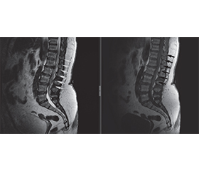
A 55-yearold female presented to our clinic with complaints of pain in the lower back radiating to the left leg. On examination, there was pain in the lumbar spine radiating to the left leg with hypostasis of the left foot. She visited a neurologist 3 weeks before, who recommended pharmacotherapeutic treatment and lumbar spine MRI. There was no relief from conservative treatment. Lumbar spine MRI done 11 days before demonstrated an L3-L4 intervertebral disc herniation caudally to the left intervertebral foramen compressing the left L4 spinal nerve and L5-S1 intervertebral disc protrusion compressing the left S1 spinal nerve root with a foraminal component (Fig. 1, 2). Due to the clinical presentation, we decided to perform a microlumbar discectomy on the L3 and L5 level on the left; the surgery lasted for 55 minutes. From patient’s anamnesis, she has hypertension, with blood pressure 180/100, heart rate 80 bpm, controlled with daily enalapril 10 mg. The patient had varicose veins of the lower extremities with occasional edema. Consultation with cardiologist and vascular surgeon revealed no contraindication to surgery (microlumbar discectomy).
The vascular surgeon recommended a lower extremity elastic bandage and elective phlebectomy. Laboratory analysis was within normal range neurologically; lower extremity muscle grade D = 5/5, S = 2/5, left foot hypostasis, positive Lasgue’s sign at 30° on the left leg. Postoperative management: 2 g of cefazolin, xefocam (NSAID) for pain. The next day after surgery, the patient was lifted and mobilized with an elastic bandage. About 6 hours after mobilization, she felt severe weakness in both extremities with muscle strength of D = 3/5, S = 2/5. Emergency lumbar spine MRI demonstrated postoperative acute spinal epidural hematoma on Th11 to L5 level, as shown in Fig. 3, necessitating emergency surgical hemilaminectomy performed on Th11-L4 spinal level, where the epidural hematoma was evacuated. Patient made recovery with muscle strength at D = 4/5, S = 3/5 on the 5th day; cefazolin 1 g, 3/7, enoxaparin 0.3, 3/7, omeprazole 20 mg, 5/7, dexamethasone 4 mg, IM 2/7, xefocam 8 mg, 5/7 were administered. Sutures were removed on the 9th postoperative day; the total in-hospital stay was 13 days.
MRI scans were performed 11 days before presentation to our hospital, showing sagittal and axial plain images of the lumbar spine, demonstrating disc herniation and protrusion at multiple levels, including L3-L4 and L5-S1. There is a left foraminal component of the herniation leading to the clinical symptoms. In addition, there is clear evidence of spondylosis, as shown in Fig. 1, 2.
As stated above, the patient stayed in the intensive care unit for 24 hours. She was given cefazolin (antibiotic), enoxaparin (blood thinner), dexamethasone (steroidal anti-inflammatory medication), and xefocam for pain relief. There was a rapid improvement in motor strength up to D = 4/5 and S = 3/5 on the 5th day. In-hospital stay were 13 days in total, then woman was transferred to a rehabilitation care.
Fig. 3 shows postoperative lumbar spine changes after discectomy on MRI (sagittal view). The mixed signal intensity in the posterior epidural space between Th11 and L5 results in considerable compression of the thecal sac and nerve roots [19]. The postoperative epidural collection demonstrated heterogeneous signal intensity. It was predominantly hyperintense with iso- and hypointense components on T2-weighted images and predominantly isointense to hypointense on T1-weighted images. This is further exacerbated by pSEH, which makes the overall condition much more complicated. There have been changes in the adjacent soft tissues after the operation, and no evidence of infection. The overall findings are consistent with the patient’s acute neurological deterioration after surgery [11].
/15.jpg)
Early rehabilitation was advantageous to the quick recovery of ambulation and balance; continuation of rehabilitation did not cause any evidence of relapses on follow-up. The case is consistent with the existing literature, which suggests that decompression therapy within 12 hours after symptom onset has the greatest chance of complete neurological recovery and functional independence [17].
The case presented above demonstrated the importance of postoperative critical care and understanding the prognosis of neurological recovery in pSEH related to rapid diagnosis and timely surgical decompression. The timely detection of acute postoperative neurological deterioration, timely imaging, and surgical intervention helped our patient recover almost completely on time [21–26]. MRI proved to be a very useful diagnostic method for confirming pSEH and ruling out other differential diagnoses.
Discussion
The case study describes a 55-year-old female patient with hypertension and varicose veins who developed pSEH from T11 to L5. In addition, a rapid bilateral weakness is observed several hours after microlumbar discectomy [3, 10]. This study highlighted several clinically important points, providing insights into the pathophysiology, diagnosis, management, and prognosis of lumbar SSEH/pSEH among hypertensive women with venous insufficiency. The methodology states the need to combine academic research and clinical models in practice, thereby making early diagnosis and interdisciplinary approaches to postoperative spinal problems more sensitive. To validate the results, data triangulation was applied; the interpretations of the reviewed literature were compared with the empirical results of the case to establish patterns and variations [1, 3]. This methodology ensured that evidence-based knowledge was complemented by first-hand clinical experience, thus helping to underscore practical implications for healthcare workers [4, 18]. The formation of epidural hematoma after microlumbar discectomy is considered to be very rare, but it can have a devastating impact. Symptomatic pSEH has a prevalence of less than 0.3 % in instrumented spine procedures, and most symptomatic pSEH develop in the early postoperative period [3, 18, 19, 22]. The first day is crucial in close observation. There are reports where spontaneous postoperative epidural hematomas have occurred, which are mainly observed in the thoracic segment of the spine, having a male dominance in multiple series [23]. Early mobilization of the patient postoperatively can be a risk factor as it increases the intrathoracoabdominal pressure; as a result, strain may occur on the internal vertebral venous plexus, which contain no valves [3, 5]. Acute alteration of position and walking can thus turn an invisible venous ooze into a clinically noticeable hematoma. Such a pathophysiologic connection provides an argument for cautious mobilization following high-risk cases with comorbidity. In addition to this, other risk factors like multilevel surgery and use of anticoagulants or coagulopathy are associated with an increased risk of bleeding, necessitating an increase in overall surveillance [8, 15].
Conclusions
In conclusion, it can be noted that symptomatic pSEH is considered to be a rare but a critical emergency case related to the neurological aspect. Out findings highlight the importance of timely MRI in a suspicious case and immediate surgical decompression in confirmed cases. Here are some critical factors that support informed decisions to achieve favorable outcomes in pSEH: early detection, early imaging, and timely hematoma evacuation, as evidenced by near-complete neurological recovery of the patient in a short time.
Received 11.04.2026
Revised 06.06.2026
Accepted 12.07.2026
1. Ahmad Z, Ahmad J, Aslam A, Ullah S, Khan U, Cole K, et al. Acute Spontaneous Spinal Epidural Hematoma in a Patient on a Direct Oral Anticoagulant (DOAC): A Case Report. Cureus. 2025 Jul 16;17(7):e88105. doi: 10.7759/cureus.88105.
2. Cheng J, Li B, Liu S, Liu Y, and Zhao Y. Case report: a patient with spontaneous spinal epidural hematoma recovered after conservative treatment. Front Med. 2025;12:1509903. doi: 10.3389/fmed.2025.1509903.
3. Epstein NE, Agulnick MA. Perspective: postoperative spinal epidural hematomas (pSEH) should be treated, not ignored. Surg Neurol Int. 2023 Oct 13;14:363. doi: 10.25259/SNI_772_2023.
4. Hsu CJ, Lin PZ, Ju DT, Hueng DY, Tseng KY. Prognostic Factors and Treatment Efficacy in Spontaneous Spinal Epidural Hematoma: A Single Center Experience and Literature Review. In Vivo. 2024 Sep-Oct;38(5):2415-2424. doi: 10.21873/invivo.13710.
5. Alexiadou-Rudolf C, Ernestus RI, Nanassis K, Lanfermann H, Klug N. Acute nontraumatic spinal epidural hematomas. An important differential diagnosis in spinal emergencies. Spine (Phila, Pa 1976). 1998 Aug 15;23(16):1810-3. doi: 10.1097/00007632-199808150-00018.
6. Lisheng H, Dong Z, Xuedong B, Jinglei S, Shaokui N, Tianjun G, et al. Successful treatment of thoracic myelopathy caused by spontaneous spinal epidural hematoma (SSEH) combined with calcification of the ligamentum flavum (CLF) by posterior percuta–neous endoscopic surgery (PPES): A case report. Front Surg. 2023 Jan 11;9:1077343. doi: 10.3389/fsurg.2022.1077343.
7. Nitta K, Imamura H, Kamijo H, Maeda Y, Uchida M, Akita M, et al. Outcomes of Conservative Management of Spontaneous Spinal Epidural Hematomas: A Retrospective Study. Cureus. 2025 Jun 5;17(6):e85430. doi: 10.7759/cureus.85430.
8. Nitta K, Imamura H, Miyama H, Mori K, Hamano Y, et al. A retrospective analysis of 30 patients with spontaneous spinal epidu–ral hematoma. Interdisciplinary Neurosurgery. 2021;25:101216. doi: 10.1016/j.inat.2021.101216.
9. Ogawa K, Akimoto T, Hara M, Fujishiro M, Uei H, Nakajima H. Two Patients with Spontaneous Spinal Epidural Hematoma Carrying a Good Prognosis without Surgical Operations. Neurol Int. 2023 Mar 7;15(1):362-370. doi: 10.3390/neurolint15010024.
10. zer M, Demirta OK. Comparison of lumbar microdiscectomy and unilateral biportal endoscopic discectomy outcomes: a single-center experience. J Neurosurg Spine. 2023 Dec 8;40(3):351-358. doi: 10.3171/2023.10.SPINE23718.
11. Peng D, Yan M, Liu T, Yang K, Ma Y, Hu X, et al. Prognostic Factors and Treatment Efficacy in Spontaneous Spinal Epidural Hematoma: A Multicenter Retrospective Study. Neurology. 2022 Aug 23;99(8):e843-e850. doi: 10.1212/WNL.0000000000200844.
12. Proulx ST. Cerebrospinal fluid outflow: a review of the historical and contemporary evidence for arachnoid villi, perineural routes, and dural lymphatics. Cell Mol Life Sci. 2021 Mar;78(6):2429-2457. doi: 10.1007/s00018-020-03706-5.
13. Stetkarova I, Ehler E, Brabec K, Jelinkova L, Chylova M, Weichet J, et al. Spontaneous spinal epidural hematoma: management and main risk factors in the era of anticoagulant/antiplatelet treatment. Neurol Neurochir Pol. 2021;55(6):574-581. doi: 10.5603/PJNNS.a2021.0066.
14. Tombeng MA, Doi K, Mahadewa TGB, Tani S, Mizuno J. Spontaneous spinal epidural hematoma in children: a case report and literature review. J Spine Surg. 2024 Sep 23;10(3):606-615. doi: 10.21037/jss-24-49.
15. Wu H, Huang X. Spontaneous spinal epidural hematoma (SSEH) after cesarean section under epidural anesthesia: A case report. Heliyon. 2023 Nov 25;9(12):e22855. doi: 10.1016/j.heliyon.2023.e22855.
16. Abu-Abaa M, Jumaah O, Mousa A, Al-Qaysi G. A Spontaneous Spinal Epidural Hematoma Secondary to Long-Term Low-Dose Aspirin and Clopidogrel Use: A Case Report. Cureus. 2023 Feb 2;15(2):e34537. doi: 10.7759/cureus.34537.
17. Perez JC, Crespo M, Velez C, Caceres J. Inpatient rehabilitation following operative spontaneous spinal epidural hematoma mimicking stroke: a case report. Spinal Cord Ser Cases. 2016 Jul 7;2:16007. doi: 10.1038/scsandc.2016.7.
18. Saitta BH, Shultz P, Hanson K, Mikhael MM. Post-Ope–rative Spinal Epidural Hematoma: Are We Discontinuing Aspirin Early Enough? Global Spine J. 2023 Oct;13(8):2255-2261. doi: 10.1177/21925682221079259.
19. Smeijers S, Collignon F, Costa E, Desmedt K, Pouleau HB, Vantomme N, et al. Consensus statement by the Belgian Society of Neurosurgery and literature review on the diagnosis and management of postoperative spinal epidural hematoma. Brain Spine. 2024 Sep 11;4:103904. doi: 10.1016/j.bas.2024.103904.
20. Kim JE, Choi DJ, Kim MC, Park EJ. Risk Factors of Post–operative Spinal Epidural Hematoma After Biportal Endoscopic Spinal Surgery. World Neurosurg. 2019 Sep;129:e324-e329. doi: 10.1016/j.wneu.2019.05.141.
21. Butler AJ, Donnally CJ 3rd, Goz V, Basques BA, Vaccaro AR, Schroeder GD. Symptomatic Postoperative Epidural Hematoma in the Lumbar Spine. Clin Spine Surg. 2022 Nov 1;35(9):354-362. doi: 10.1097/BSD.0000000000001278.
22. Chen Q, Zhong X, Liu W, Wong C, He Q, Chen Y. Incidence of postoperative symptomatic spinal epidural hematoma requiring surgical evacuation: a systematic review and meta-analysis. Eur Spine J. 2022 Dec;31(12):3274-3285. doi: 10.1007/s00586-022-07421-6.
23. Glotzbecker MP, Bono CM, Wood KB, Harris MB. Postoperative spinal epidural hematoma: a systematic review. Spine (Phila, Pa 1976). 2010 May 1;35(10):E413-20. doi: 10.1097/BRS.0b013e3181d9bb77.
24. Robinson L, Forget P, Nesvadba D. Systemic Hypertension and Postoperative Symptomatic Spinal Epidural Hematoma: A Sco–ping Review. Journal of Neurosurgical Anesthesiology. 2024;36(4):303-308. doi: 10.1097/ANA.0000000000000947.
25. Djurasovic M, Campion C, Dimar JR 2nd, Glassman SD, Gum JL. Postoperative Epidural Hematoma. Orthop Clin North Am. 2022 Jan;53(1):113-121. doi: 10.1016/j.ocl.2021.08.006.
26. Leroy HA, Portella T, Amouyel T, Bougeard R, Assaker R, Mourier KL. Management of symptomatic postoperative epidu–ral hematoma in spine surgery: medicolegal implications. Orthop Traumatol Surg Res. 2021 Nov;107(7):103024. doi: 10.1016/j.otsr.2021.103024.