
Oral and General Health Том 7, №3, 2026
Вернуться к номеру
Комп’ютерна томографія у випадках із застосуванням додаткового методу лікування дисфункції скронево-нижньощелепного суглоба
Авторы: Мірза Р.О., Лихота К.М.
Національний університет охорони здоров’я України імені П.Л. Шупика, м. Київ, Україна
Рубрики: Стоматология
Разделы: Клинические исследования
Версия для печати
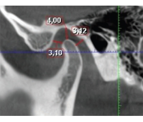
Актуальність. Співвідношення щелеп, що є рекомендованим для подальшої оклюзійної реабілітації, набуває статусу лікувального за умови відсутності клінічних проявів патології скронево-нижньощелепного суглоба і жувальних м’язів. У терапевтичному співвідношенні щелеп методи візуалізації скронево-нижньощелепного суглоба найчастіше застосовуються для оцінки змін у співвідношенні між суглобовою головкою і суглобовим диском, дегенеративних проявів стану кісткової тканини. На рівні аналізу комп’ютерних томограм показники норми для лікувального співвідношення щелеп можуть бути індивідуалізованими, як і терміни адаптації жувальної системи під час неінвазивного лікування патології скронево-нижньощелепного суглоба. Це твердження обґрунтовано тим, що в певних випадках відсутність клінічних ознак патології скронево-нижньощелепного суглоба і жувальних м’язів виявлено при дистальному, центральному і передньому положенні суглобової головки в суглобовій ямці. Мета: визначити особливості адаптації жувальної системи за терапевтичного співвідношення щелеп, виявити оборотні ускладнення, що виникають під час неінвазивного лікування патології скронево-нижньощелепного суглоба, та оцінити можливості їх усунення. Матеріали та методи. Виконано клінічне обстеження 275 (100 %) пацієнтів віком від 18 до 35 років, яким в подальшому було запропоновано пройти оклюзійну реабілітацію, що включала вимірювання відстані між різцями при найбільшому непримусовому відкриванні рота, пальпацію скронево-нижньощелепного суглоба і жувальних м’язів, встановлення попереднього діагнозу відповідно до міжнародного протоколу обстеження DC/TMJ. Проаналізовано 275 комп’ютерних томограм для правої і лівої сторони до та після неінвазивного лікування патології скронево-нижньощелепного суглоба і жувальних м’язів, що включало застосування гнатологічної капи, оклюзійних накладок. На комп’ютерних томограмах вимірювались передня, верхня і задня суглобові щілини на стороні виявлення клінічних ознак дисфункції скронево-нижньощелепного суглоба, асоційованої з положенням диска без обмеженого відкривання рота, клінічних і рентгенологічних ознак остеоартрозу. Додаткове лікування дисфункції скронево-нижньощелепного суглоба реалізувалось шляхом застосування електростимуляції жувальних м’язів біполярним імпульсним струмом 24–120 Гц. Результати. Під час неінвазивного лікування 223 пацієнтів з клінічними ознаками зміщення диска з репозицією за класифікацією DC/TMJ виявлено прояви спазму та скутості в жувальних м’язах при відкриванні рота, що виникли після застосування оклюзійних накладок у 3 (1,3 %) пацієнтів, і обмежене відкривання рота, що з’явилось в перші дні користування гнатологічною шиною додатково у 3 (1,3 %) обстежених. За терапевтичного співвідношення щелеп, підтвердженого клінічним обстеженням і комп’ютерною томографією, у 6 (2,6 %) пацієнтів вдалося повністю усунути спазм жувальних м’язів та обмеження відкривання рота за допомогою електростимуляції жувальних м’язів тривалістю 7–15 хв. У 6 (2,6 %) обстежених під час використання оклюзійних накладок за терапевтичного співвідношення щелеп виникли ускладнення, що клінічно проявлялися спазмом жувальних м’язів. Крім того, у перші дні активного використання гнатологічної шини виявлено зміщення суглобового диска з репозицією та періодичним блокуванням рухів нижньої щелепи. З’ясовано, що в період неінвазивного лікування дисфункції скронево-нижньощелепного суглоба у 6 (2,6 %) обстежених електростимуляція жувальних м’язів здатна забезпечити фізіологічну адаптацію жувальної системи. Отже, у пацієнтів із клінічними та рентгенологічними ознаками остеоартрозу скронево-нижньощелепного суглоба під час неінвазивного лікування зміщення суглобового диска з репозицією не виявлено жодного випадку оборотних ускладнень. Висновки. При неінвазивному лікуванні дисфункції скронево-нижньощелепного суглоба, асоційованої з положенням диска, що має ознаки репозиції, зберігається ризик виникнення клінічних проявів спазму жувальних м’язів і обмеженого відкривання рота при центральному і передньому положенні суглобової головки в суглобовій ямці, визначеному на комп’ютерних томограмах. Такий вид адаптації жувальної системи є підставою до застосування електростимуляції поверхневих жувальних м’язів з метою скорочення термінів лікування. За неінвазивного лікування патології скронево-нижньощелепного суглоба, асоційованої зі зміщенням суглобового диска з репозицією, ризик виникнення спазму жувальних м’язів і раптового обмеження відкривання рота за терапевтичного співвідношення щелеп є мінімальним у пацієнтів із клінічними та рентгенологічними ознаками остеоартрозу, підтвердженими даними комп’ютерної томографії.
Background. The jaw relationship recommended for subsequent occlusal rehabilitation may be considered therapeutic provided that there are no clinical signs of pathology of the temporomandibular joint (TMJ) or masticatory muscles. In the context of therapeutic jaw relationships, methods of TMJ imaging are most commonly used to assess changes in the relationship between the mandibular condyle and the articular disc, as well as degenerative changes in the osseous structures. Analysis of computed tomography (CT) images suggests that normative parameters for a therapeutic jaw relationship may be individualized, as may the duration of the masticatory system adaptation during non-invasive treatment for TMJ pathology. This assumption is based on the fact that in certain cases, the absence of clinical signs of pathology of the TMJ and masticatory muscles was observed with the mandibular condyle positioned distally, centrally, or anteriorly within the glenoid fossa. Aim: to determine the characteristics of masticatory system adaptation occurring in a therapeutic jaw relationship and to identify reversible complications arising during the non-invasive treatment for TMJ disorders, followed by an assessment of the feasibility of overcoming these complications. Materials and methods. Clinical examination was performed in 275 patients (100 %) aged 18–35 years who were subsequently offered occlusal rehabilitation. The examination included measurement of the interincisal distance at maximum unforced mouth opening, palpation of the TMJ and masticatory muscles, and establishment of a preliminary diagnosis based on the international Diagnostic Criteria for Temporomandibular Disorders (DC/TMD) examination protocol. A total of 275 CT scans of the right and left sides were analyzed before and after non-invasive treatment for TMJ and masticatory muscle disorders, which included the use of a gnathological splint and occlusal overlays. On CT images, the anterior, superior, and posterior joint spaces were measured on the side exhibiting clinical signs of TMJ dysfunction associated with disc positioning without limited mouth opening, as well as clinical and radiographic signs of osteoarthritis. Additional treatment for TMJ dysfunction was performed using electrical stimulation of the masticatory muscles with bipolar pulsed current at frequencies ranging from 24 to 120 Hz. Results. During non-invasive treatment of 223 patients with clinical signs of disc displacement with reduction according to the DC/TMD classification, manifestations of masticatory muscle spasm and stiffness during mouth opening were observed in 3 patients (1.3 %) following the use of occlusal overlays. Limited mouth opening developed during the first days of using a gnathological splint in an additional 3 patients (1.3 %). In the therapeutic jaw relationship verified by clinical examination and CT, the manifestations of masticatory muscle spasm and limited mouth opening were completely eliminated in 6 patients (2.6 %) by electrical stimulation of the masticatory muscles for 7–15 minutes. In 6 patients (2.6 %), clinical manifestations consistent with masticatory muscle spasm developed while occlusal overlays were used in the therapeutic jaw relationship. These patients had disc displacement with reduction accompanied by intermittent locking of mandibular movements, which became evident during the first days of active use of a gnathological splint. It was found that with non-invasive treatment for TMJ dysfunction, electrical stimulation of the masticatory muscles in 6 patients (2.6 %) facilitated a physiological adaptation of the masticatory system. Thus, no cases of reversible complications were identified in non-invasive treatment for disc displacement with reduction in patients with radiographic and clinical signs of TMJ osteoarthritis. Conclusions. During non-invasive treatment for TMJ dysfunction associated with disc positioning with reduction, there remains a risk of developing clinical manifestations of masticatory muscle spasm and limited mouth opening when the mandibular condyle is positioned centrally or anteriorly within the glenoid fossa, as determined by CT. This type of masticatory system adaptation may constitute an indication for the use of electrical stimulation of the superficial masticatory muscles to facilitate adaptation and shorten the treatment period. During non-invasive treatment for TMJ pathology associated with disc displacement with reduction, the risk of developing masticatory muscle spasm and spontaneous limitation of mouth opening is minimal with a therapeutic jaw relationship in patients with clinical and radiographic signs of osteoarthritis visualized on CT.
скронево-нижньощелепний суглоб; електростимуляція жувальних м’язів; комп’ютерна томографія
temporomandibular joint; electrical stimulation of the masticatory muscles; computed tomography