
Oral and General Health Том 7, №3, 2026
Вернуться к номеру
Функціональний стан пульпи зубів у динаміці традиційного роторного та повітряно-абразивного препарування
Авторы: Лахтін Ю.В., Кузенко Є.В., Москаленко П.О., Галич Л.В.
Сумський державний університет, м. Суми, Україна
Рубрики: Стоматология
Разделы: Справочник специалиста
Версия для печати
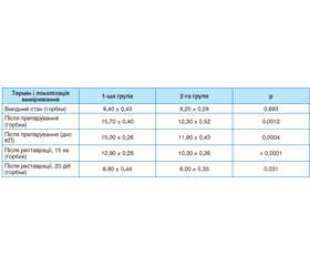
Актуальність. Збереження функціонального стану пульпи є одним із біологічних критеріїв ефективності оперативного лікування карієсу. Різні механізми роторного та повітряно-абразивного препарування можуть зумовлювати відмінності ранньої функціональної реакції пульпи на втручання. Мета: оцінити функціональний стан пульпи зубів за допомогою електроодонтометрії у динаміці традиційного роторного та повітряно-абразивного препарування каріозних порожнин. Матеріали та методи. У клінічному дослідженні з дизайном «розділеного рота» обстежено 10 пацієнтів із симетричними каріозними порожнинами I класу за Блеком (ICDAS 5 та 6) у контралатеральних нижніх молярах. Загалом досліджено 20 зубів: 10 препарували традиційним роторним методом (1-ша група), 10 — повітряно-абразивним методом (2-га група). Електроодонтометрію проводили до препарування, безпосередньо після нього, через 15 хв після завершення реставрації та через 20 діб. Для статистичного аналізу застосовували методи для парних спостережень та повторних вимірювань. Результати. Вихідні показники електроодонтометрії контралатеральних зубів були порівнянними. Безпосередньо після препарування поріг електричної відповіді пульпи підвищувався в обох групах, однак після роторного препарування він був вищим, ніж після повітряної абразії, як при вимірюванні з горбків (15,70 ± 0,40 проти 12,30 ± 0,52 мкА; p = 0,0012), так і з дна каріозної порожнини (15,00 ± 0,26 проти 11,90 ± 0,43 мкА; p = 0,0004). Через 15 хв після завершення реставрації показники знижувалися в обох групах, проте міжгрупова відмінність зберігалася (p < 0,0001). Через 20 діб спостерігалося подальше зниження порога електричної відповіді до 6,80 ± 0,44 мкА в 1-й групі та 6,00 ± 0,33 мкА в 2-й (p = 0,031); в обох групах ці показники були нижчими за вихідні. Внутрішньогрупова динаміка протягом періоду спостереження була статистично значущою в обох групах (p < 0,001). Висновки. Роторне та повітряно-абразивне препарування каріозних порожнин супроводжується часовими змінами електричної реактивності пульпи. При повітряно-абразивному препаруванні рання функціональна реакція пульпи є менш вираженою, ніж при традиційному роторному препаруванні; надалі після обох способів препарування спостерігається послідовна зміна її електричної реактивності після завершення реставраційного лікування.
Background. Preservation of pulp function is an important biological consideration in operative caries treatment. Rotary (RP) and air-abrasion cavity preparation (AAP) involve different mechanisms of hard-tissue removal and may therefore elicit different early functional responses of the dental pulp. This study aimed to assess changes in pulp functional status using electric pulp testing (EPT) during and after conventional rotary and air-abrasion preparation of carious cavities. Materials and methods. A split-mouth clinical study included 10 patients with symmetrical Black class I carious lesions (ICDAS 5–6) in contralateral mandibular molars. Twenty teeth were examined: 10 underwent conventional RP (group 1) and 10 AAP (group 2). EPT was performed before cavity preparation, immediately after it, 15 min after completion of the restoration, and at the 20-day follow-up. Statistical analysis accounted for paired observations and repeated measurements. Results. Baseline EPT values were comparable between contralateral teeth. Immediately after cavity preparation, the threshold for electrical pulp response increased with both techniques but was significantly higher after RP than after AAP, both at the cusp (15.70 ± 0.40 vs 12.30 ± 0.52 μA; p = 0.0012) and at the cavity floor (15.00 ± 0.26 vs 11.90 ± 0.43 μA; p = 0.0004). Fifteen minutes after restoration, EPT thresholds decreased in both groups, while the between-group difference remained significant (p < 0.0001). At 20 days, a further decrease was observed, with thresholds of 6.80 ± 0.44 μA in group 1 and 6.00 ± 0.33 μA in group 2 (p = 0.031); values in both groups were below their respective baseline levels. Within-group changes over the observation period were statistically significant for both preparation techniques (p < 0.001). Conclusions. Rotary and air-abrasion cavity preparation were associated with time-dependent changes in the electrical responsiveness of the dental pulp. The early functional response was less pronounced following air-abrasion than conventional rotary preparation. With both techniques, pulp electrical responsiveness continued to change after completion of restorative treatment.
зуби; карієс; препарування; повітряна абразія; роторне препарування; електроодонтометрія
teeth; dental caries; cavity preparation; air abrasion; rotary preparation; electric pulp testing