Международный эндокринологический журнал Том 17, №8, 2021
Вернуться к номеру
Ліпоїдний некробіоз: рідкісний клінічний та патоморфологічний випадок
Авторы: Ніжинська-Астапенко З.П. (1), Власенко М.В. (1), Вернигородський В.С. (1), Холод Л.П. (2), Шведка О.В. (2)
(1) — Вінницький національний медичний університет ім. М.І. Пирогова, м. Вінниця, Україна
(2) — КНП «Вінницьке обласне патологоанатомічне бюро Вінницької обласної ради», м. Вінниця, Україна
Рубрики: Эндокринология
Разделы: Справочник специалиста
Версия для печати
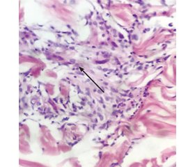
Згідно із сучасними науковими дослідженнями, ліпоїдний некробіоз (ЛН) — це захворювання, що характеризується вогнищевою дезорганізацією та ліпідною дистрофією колагену. Вважають, що в основі шкірних змін при цьому дерматозі лежить діабетична мікроангіопатія, яка супроводжується склерозуванням та облітерацією кровоносних судин, що призводить до некробіозу дерми з подальшим відкладанням у ній ліпідів. Цю патологію реєструють порівняно рідко, у середньому в 1 % хворих на цукровий діабет (ЦД). Поєднання ЛН із ЦД, за даними літератури, становить у межах від 25 до 70 %; частіше (у 40–60 % випадків) ЦД передує ЛН, а у 10–25 % випадків вони виникають одночасно. До того ж у 10–50 % випадків ЛН діагностується в осіб без супутнього ЦД. Варіативність клінічних, епідеміологічних особливостей та відносна незначна поширеність цієї патології досить часто є приводом помилкового чи пізнього діагнозу. Описаний клінічний випадок типовий стосовно епідеміологічних даних: стать, вік, наявність ЦД. Одночасно він є рідкісним щодо клінічної картини: не є класично діабетичним за локалізацією (типовими є симетричні ділянки гомілок), за зовнішнім виглядом ділянок некробіозу — гранулематозний тип некробіозу у вигляді кільцеподібної гранульоми, за гістологічною будовою — ділянка хронічного периваскулярного лімфоплазмоцитарного запалення за участі поодиноких гігантських клітин, що потребувало додаткових клінічних та анамнестичних даних для об’єктивного висновку патоморфолога. Біопсія в даному випадку використовувалась для диференціальної діагностики між кільцеподібною гранульомою та некробіотичною некрогранульомою. До того ж цей метод діагностики відіграв додаткову роль у лікуванні. Даний випадок продемонстрував, можливо, активацію клітинної та гуморальної імунної відповіді в зоні хронічного запалення у відповідь на механічне пошкодження та завершення запалення з повним відновленням тканини.
According to modern scientific researches, necrobiosis lipoidica (NL) is a disease characterized by focal disorganization and lipid collagen dystrophy. It is believed that the basis of skin changes in this dermatosis is diabetic microangiopathy that is accompanied by sclerosis and obliteration of blood vessels, which leads to necrobiosis with subsequent deposition of lipids in the dermis. This pathology is registered relatively rarely, in 1 % of patients with diabetes mellitus (DM) on average. The combination of NL with DM, according to the literature data, ranges from 25 to 70 %; more often (in 40–60 % of cases) DM is preceded by NL, and in 10–25 % of cases they occur simultaneously. In addition, in 10–50 % of cases NL is diagnosed in people without concomitant diabetes. The variability of clinical, epidemiological features and the relatively low prevalence of this pathology is often the cause for misdiagnosis or late diagnosis. The described clinical case is typical in terms of the epidemiological data: sex, age, presence of DM. At the same time, it is rare in terms of the clinical picture: it is not classically diabetic by localization (symmetrical areas of the legs are typical), by appearance of necrobiosis areas — granulomatous type of necrobiosis in the form of granuloma annulare, by histological structure — area of chronic perivascular lymphoplasmocytic inflammation with the involvement of single giant cells, which required additional clinical and anamnestic data for an objective report of the pathologist. Biopsy in this case was used as a differential diagnosis between granuloma annulare and necrobiotic necrogranuloma. In addition, this method of diagnosis has played an additional therapeutic role. This case may have demonstrated the activation of the cellular and humoral immune response in the area of chronic inflammation in response to a mechanical damage and the resolution of inflammation with complete tissue repair.
цукровий діабет; ліпоїдний некробіоз; диференціальна діагностика; лікування
diabetes mellitus; necrobiosis lipoidica; differential diagnosis; treatment
Вступ
Клінічний випадок
Обговорення
Висновки
- Feily A., Mehraban S. Treatment Modalities of Necrobiosis Lipoidica: A Concise Systematic Review. Dermatol. Reports. 2015, Jun 8. 7(2). 5749. doi: 10.4081/dr.2015.5749. PMID: 26236446; PMCID: PMC4500868.
- Semenova D.A., Tokmakova A.Yu. Necrobiosis lipoidica in diabetic patients: pathogenetic and clinical features. Diabetes mellitus. 2011. 14(4). 51-54. https://doi.org/10.14341/2072-0351-5817. (In Russian)
- Mistry B.D., Alavi A., Ali S., Mistry N. A systematic review of the relationship between glycemic control and necrobiosis lipoidica diabeticorum in patients with diabetes mellitus. Int. J. Dermatol. 2017 Dec. 56(12). 1319-1327. doi: 10.1111/ijd.13610.
- Fehlman J.A., Burkemper N.M., Missall T.A. Ulcerative necrobiosis lipoidica in the setting of anti-tumor necrosis factor-α and hydroxychloroquine treatment for rheumatoid arthritis. JAAD Case Rep. 2017, Mar 12. 3(2). 127-130. doi: 10.1016/j.jdcr.2017.01.006.
- Reid S.D., Ladizinski B., Lee K., Baibergenova A., Alavi A. Update on necrobiosis lipoidica: a review of etiology, diagnosis, and treatment options. J. Am. Acad. Dermatol. 2013 Nov. 69(5). 783-791. doi: 10.1016/j.jaad.2013.05.034.
- Hammer E., Lilienthal E., Hofer S.E., Schulz S., Bollow E., Holl R.W. DPV Initiative and the German BMBF Competence Network for Diabetes Mellitus. Risk factors for necrobiosis lipoidica in Type 1 diabetes mellitus. Diabet Med. 2017 Jan. 34(1). 86-92. doi: 10.1111/dme.13138.
- Erfurt-Berge C., Dissemond J., Schwede K., Seitz A.T., Al Ghazal P., Wollina U., Renner R. Updated results of 100 patients on clinical features and therapeutic options in necrobiosis lipoidica in a retrospective multicentre study. Eur. J. Dermatol. 2015 Nov-Dec. 25(6). 595-601. doi: 10.1684/ejd.2015.2636. PMID: 26575980.
- Hashemi D.A., Nelson C.A., Elenitsas R., Rosenbach M. An atypical case of papular necrobiosis lipoidica masquerading as sarcoidosis. JAAD Case Rep. 2018, Sep 14. 4(8). 802-804. doi: 10.1016/j.jdcr.2018.07.011. PMID: 30246132; PMCID: PMC6141674.
- Pourang A., Sivamani R.K. Treatment-resistant ulcerative necrobiosis lipoidica in a diabetic patient responsive to ustekinumab. Dermatol. Online J. 2019, Aug 15. 25(8). 13030/qt2q05z4rw. PMID: 31553866.


/75.jpg)
/76.jpg)