Інформація призначена тільки для фахівців сфери охорони здоров'я, осіб,
які мають вищу або середню спеціальну медичну освіту.

Підтвердіть, що Ви є фахівцем у сфері охорони здоров'я.


Коморбідний ендокринологічний пацієнт

Коморбідний ендокринологічний пацієнт

Международный эндокринологический журнал Том 18, №2, 2022

Вернуться к номеру

Гіперкортицизм на тлі реабілітації після COVID-19 (клінічний випадок)

Авторы: Kravchenko V. (1), Rakov O. (1), Slipachuk L.V. (2)
(1) — State Institution “V.P. Komisarenko Institute of Endocrinology and Metabolism of the NAMS of Ukraine”, Kyiv, Ukraine
(2) — Bohomolets National Medical University, Kyiv, Ukraine

Рубрики: Эндокринология

Разделы: Справочник специалиста

Версия для печати


Резюме

Ураження легеневої тканини належить до переважних ускладнень вірусного захворювання COVID-19. Також описані ускладнення з боку інших органів, у тому числі з боку ендокринних залоз. Повідомляється про ураження надниркових залоз зі зниженням їх функції на тлі COVID-19. Декілька досліджень автопсії надниркових залоз у пацієнтів виявили судинний тромбоз, ліпідну дегенерацію коркового шару, ішемічний некроз, паренхіматозні інфаркти та інші ураження, що призводять до зниження функції надниркових залоз. Також можливий центральний механізм дисфункції надниркових залоз унаслідок крововиливу та некрозу гіпофіза. У статті подано рідкісний випадок розвитку гіперкортицизму у молодої жінки після одужання від COVID-19. Зважаючи на високий рівень АКТГ (157 і 122 пг/мл), негативний нічний дексаметазоновий тесту і високі 24-годинні показники екскреції вільного (добового) кортизолу з сечею, автори попередньо запідозрили хворобу Кушинга. Хромогранін А був у межах норми — 21,35 (референтне значення < 100). Інші тести показали підвищений рівень дигідротестостерону — 780,6 пг/мл (референтні значення 24–368 пг/мл). Рівні інших досліджених гормонів передньої частки гіпофіза були в межах норми. Згідно з клінічними рекомендаціями, препаратом вибору для короткочасного лікування цього захворювання є інгібітори стероїдогенезу — кетоконазол. Ефективність такої схеми лікування раніше була доведена іншими дослідниками. У нашому випадку призначали кетоконазол у дозі 400 мг 2 рази на добу та каберголін (достинекс) у початковій дозі 1 мг на добу. Зважаючи на низький рівень вітаміну D у сироватці крові, рекомендовано продовжувати прийом вітаміну D у дозі 4000 МО на добу. Рекомендовано через 2 місяці контролювати лабораторні показники крові — кортизол сироватки, АКТГ, АСТ, АЛТ, електроліти, 25(OH)D, рівень глюкози в крові та визначитися з подальшою тактикою ведення хворої.

Damage to the lung tissue is a predominant complication of the viral disease COVID-19. Recently, there have been complications from other organs, including highly vascularized endocrine glands. Regarding the adrenal glands, there are reports of their damage with a decrease in their function. Сhanging the function of the adrenal glands (AG) in patients with or after COVID-19 is important. A few adrenal autopsy studies in patients have revealed vascular thrombosis, cortical lipid degeneration, ischemic necrosis, parenchymal infarcts, and other lesions leading to a decrease in AG function. The central mechanism of adrenal dysfunction through hemorrhage and necrosis of the pituitary gland is also possible. This paper presents a rare case of the development of hypercortisolism in a young woman after recovering from COVID-19. Based on high ACTH levels (157 and 122 pg/ml), a negative nocturnal dexamethasone test, and high 24-hour urinary free (daily) cortisol excretion rates, we tentatively suspected Cushing’s disease. Chromogranin A was within the normal range of 21.35 (reference value < 100). Other tests showed an elevated dihydrotestosterone level of 780.6 pg/ml (reference values 24–368 pg/ml). The levels of other anterior pituitary hormones tested were within the normal range. According to clinical guidelines, the drug of choice for the short-term treatment of this disease is steroidogenesis inhibitors — ketoconazole. The effectiveness of such a treatment regimen was previously brought to light by others. In our case, ketoconazole was prescribed at a dose of 400 mg 2 times a day and cabergoline (dostinex) at an initial dose of 1 mg per day. Given the low levels of vitamin D in the blood serum, it was recommended to continue taking vitamin D at a dose of 4000 IU per day. It was recommended to control blood laboratory parameters — serum cortisol, ACTH, AST, ALT, electrolytes, 25(OH)D, blood glucose level after 2 months and decide on further tactics for managing the patient.


Ключевые слова

синдром Кушинга, гіперкортицизм, COVID-19, лікування, клінічний випадок

Cushing’s syndrome; hypercortisolism; COVID-19; treatment; case report

Abbrevations

ACTH — adrenocorticotropic hormone; CS — Cushing’s syndrome; AG — adrenal glands; PCR-test — Polymerase Chain Reaction; ODT — overnight dexamethasone test; AST — aspartate aminotransferase; ALT — alanine aminotransferase; 25-(OH)D — vitamin D.

Introduction

The COVID-19 pandemic continues. To date, about 330 million people have been infected with COVID and 5.5 million have died in the world. Millions of new cases registered every day [1]. The main consequence of the disease is acute respiratory syndrome with the development of pneumonia and complications from the cardiovascular system, kidneys and other organs and tissues including vasculitis, arteriolar and venous thrombosis, hypoxic cell injury, subsequent immune response and cytokine storm [2, 3]. This effect can manifest itself in highly vascularized organs, including endocrine glands. The attention of specialists directs to the study and treatment of diabetes mellitus and thyroid diseases. Diabetes is an important risk factor for poor prognosis of COVID-19 disease and mortality [4, 5]. Damage to the thyroid gland complicates the disease with its insufficiency or hyperfunction [6–8]. A significant place in the treatment of COVID-19 belongs to glucocorticoids [9–11]. 
Therefore, changing the function of the adrenal glands (AG) in patients with or after COVID-19 is important. A few adrenal autopsy studies in patients have revealed vascular thrombosis, cortical lipid degeneration, ischemic necrosis, parenchymal infarcts, and other lesions leading to a decrease in AG function [12–15]. The central mechanism of adrenal dysfunction through hemorrhage and necrosis of the pituitary gland is also possible [12, 16]. The main manifestation of the opposite state of “hypercortisolism” is Cushing’s syndrome (CS). The prevalence of CS is about 40 cases per million, and the estimated incidence is 0.7–2.4 cases per million per year [17]. Reports of COVID-19 infection in patients with CS are rare, and the treatment of such patients needs a special approach [18]. An analysis of the literature available to us over the past two years has shown that there is no description of cases of the occurrence of endogenous hypercortisolism against the background of coronavirus disease. In this publication, we present a description of a patient who first developed ACTH-dependent hypercortisolism after recovering from mild COVID-19.

Case description

The female T., European race, unmarried, age 22 years, height 162 cm, weight 80 kg, first applied to the consultative polyclinic of the institute for the study of endocrinologist with complaints of weight gain (approximately 20 kg) over the past 8 months. There were also complaints of recurrent headaches, general weakness, fatigue, rises in blood pressure (BP), the presence of stretch marks in the anterior wall of the abdomen, hips, chest, menstrual irregularities (like oligomenorrhea), a feeling of internal discomfort, anxiety, shortness of breath with little exertion. From the anamnesis of the disease, it is known that at the end of February 2021, the patient lost her sense of smell, subfebrile body temperature (37–37.5), general weakness appeared. The family doctor examined the patient at the place of residence. PCR-test was positive, confirming SARS-CоV-2. She treated on an outpatient basis, as the disease proceeded in a mild form. A month after the appearance, she noticed the appearance of single purple furrows on the skin of the abdomen, on the surface of the inner thighs, she began to gain weight sharply, headaches and shortness of breath appeared during physical exertion. When used by an endocrinologist, an elevated serum cortisol of 26.4 μg/dl (4.3–22.4) was detected, and ACTH levels were within the reference values. Before contacting the institute’s polyclinic, the patient consulted doctors of various specialties who diagnosed hypercortisolinemia against the background of diencephalic dysfunction. The patient took antihypertensive, antidiuretic drugs, as well as drugs that improve cerebral circulation. The general condition continued to deteriorate, weight increased, menstrual irregularities appeared, appearance changed (the face became puffy, edematous), trophic manifestations on the skin of the abdomen, shoulders, chest progressed, muscle weakness increased. In view of which, patient was referred to the clinic of the Institute of Endocrinology for diagnosis.
When studying the anamnesis, it became known that both parents were Ukrainians by nationality, the marriage was not consanguineous. Pedigree is not burdened. She is the second child in the family. The mother’s pregnancy proceeded without any peculiarities and ended in natural delivery. Birth weight — 4200 g, height — 54 cm, see Apgar score — 7/9 points. 
The patient denied of taking any preparations containing glucocorticoids, illegal drug use. Heredity for endocrine diseases is not burdened. There was no allergic reaction to medication.
Clinically, the patient had Cushingoid facial features, abdominal obesity with significant thinning of the lower extremities with severe proximal myopathy. On the front surface of the abdomen, the inner surface of the shoulders, the hips are wide (more than 4 cm) crimson-red, purple stripes of stretching (furrows) (fig. 1).
The described changes were accompanied by an increase in blood pressure up to 150/110 mm Hg, and body mass index (BMI) up to 30.5 kg/m2.
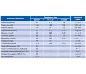
General blood analysis revealed no abnormalities in the content of basic elements. Alanine aminotransferase and aspartate aminotransferase contents were elevated to 60 and 56 U/l with reference values (4–41) and (4–37), respectively. This indicated impaired liver function. There was also a reduced content of hydroxyvitamin D3 — 24.8 nmol/l, with the required norm of > 75 nmol/l. According to the results of glycosylated hemoglobin, there were no violations of carbohydrate metabolism (table 1).
As can be seen from the table 2, an increase in the basal level of ACTH 157.0 pg/ml attracts attention (the norm is 7.2–63.30 pg/ml). All repeat ACTH measurements were elevated (111 pg/ml, 122 pg/ml, respectively). The basal serum cortisol level was 96.14 µg/dl (morning 7 to 9 am: 4.3–22.4). The patient’s primary (overnight) test to confirm the presence of endogenous hypercorticism (this is a suppression test of 1 mg dexamethasone, i.e. 2 tablets of 0.5 mg dexamethasone) was negative in the patient. We applied 2 tablets of dexamethasone 0.5 mg each, the suppression of serum cortisol levels did not occur properly. The pre-test blood cortisol level was 96.14 mcg/dl, and after 2 dexamethasone tablets taken at 23:00 hours, the blood cortisol level was 50.0 and 78,1 mcg/dl (reference values — 4,3–22.4 mcg/dl). Initial studies showed that 24-hour free urine cortisol was more than 716 mcg, which is 2 times the reference values. High levels of free blood cortisol persisted at the second and third studies (511 and 619 mcg/24 hours), respectively.
Based on high ACTH levels (157–111 and 122 pg/ml, respectively), a negative nocturnal dexamethasone test, and high 24-hour urinary free (daily) cortisol excretion rates, we tentatively suspected Cushing’s disease. Chromogranin A was within the normal range of 21.35 (reference value < 100). Other tests showed an elevated dihydrotestosterone level of 780.6 pg/ml (reference values 24–368 pg/ml). The levels of other anterior pituitary hormones tested were within the normal range.
The patient’s visit to the Institute of Endocrinology coincided with another lockdown due to the global coronavirus pandemic, due to which the work of specialized hospitals was suspended or extremely limited, and the patient was not hospitalized.
According to clinical guidelines, the drug of choice for the short-term treatment of this disease is steroidogenesis inhibitors — ketoconazole [20]. The effectiveness of such a treatment regimen was previously brought to light by others. In our case, ketoconazole was prescribed at a dose of 400 mg 2 times a day and cabergoline (dostinex) at an initial dose of 1 mg per day. Given the low levels of vitamin D in the blood serum (table 1), it was recommended to continue taking vitamin D at a dose of 4000 IU per day. It was recommended to control blood laboratory parameters — serum cortisol, ACTH, AST, ALT, electrolytes, 25(OH)D, blood glucose level after 2 months and decide on further tactics for managing the patient.

Discussion

CS can be exogenous as a result of chronic corticosteroid use or endogenous due to overproduction of cortisol by the adrenal glands. In our case, with coronavirus disease, the patient did not take corticosteroids. In about 80 % of cases, endogenous CS is caused by overstimulation of the adrenal glands due to abnormally elevated levels of ACTH, due to an ACTH-secreting pituitary tumor (Cushing’s disease) or an extrapituitary ACTH-secreting tumor (ectopic ACTH). Difficulties in identifying the primary focus of ACTH hypersecretion may be due to its small size, the absence or non-specificity of the clinical picture from the primary focus, and early metastasis in the case of malignant tumors. In addition, the clinical manifestations of hypercorticism can also be considered as independent nosological forms, which creates certain difficulties at the stages of early diagnosis and timely treatment of ACTH-dependent hypercorticism. In our case, the content of ACTH in the blood was significantly increased. However, magnetic resonance imaging of the pituitary showed no obvious adenoma. Multislice computed tomography of the neck, chest and abdominal organs, retroperitoneal space did not reveal an extrapituitary ectopic tumor.
In about 20 % of cases, CS is associated with autonomous, unregulated adrenal cortisol secretion [21, 22]. The condition of the patient we observed was characterized by the presence of a detailed clinical picture of hypercortisolism (burgundy striae, ”bruises“ on the skin, rounding and reddening of the face against the background of weight gain, muscle weakness, increased blood pressure) in the absence of a source of ACTH-ectopia, despite a thorough diagnostic search. Elevated levels of ACTH in the blood during follow-up, as well as negative ODT with 1 mg of dexamethasone, high levels of cortisol in daily urine and saliva in the evening, indicated ACTH-dependent Cushing’s syndrome. This case shows that the diagnosis of ACTH-dependent hypercortisolism, the search for the primary focus, even with the use of modern imaging diagnostic methods, present considerable difficulties and require long-term follow-up and further topical search. Clinical observations by other investigators suggest that the hypothalamic-pituitary-adrenal (HPA) axis appears to be directly involved in SARS-CoV or indirectly due to virus-induced hypophysitis [23].
Medical therapy during the COVID-19 pandemic may represent an interesting and effective approach for patients with CS, as it can rapidly lower circulating cortisol levels and therefore improve clinical comorbidities with a potential impact on the course of COVID-19 infection. In our case, we were treated with ketoconazole (a powerful inhibitor of steroidogenesis 400 mg twice a day, and with carbegoline (dostinex), which is a D2-receptor agonist, and also continued monitoring of the disease progression. Both drugs are present on the Ukrainian market. 

Conclusions

COVID-19 can cause damage in the pituitary-adrenal system with the onset of Cushing’s syndrome and requires careful approaches for diagnosis and treatment.
 
Received 04.03.2022
Revised 17.03.2022
Accepted 27.03.2022

Список литературы

  1. WHO. Weekly operational update on COVID-19 — 22 February 2022. https://www.who.int/publications/m/item/weekly-operational-update-on-covid-19-22-february-2022.
  2. Ding Y., Wang H., Shen H., Li Z., Geng J., Han H., Cai J., et al. The clinical pathology of severe acute respiratory syndrome (SARS): a report from China. J. Pathol. 2003. 200. 282-289. doi: 10.1002/path.1440. 
  3. Guo Y., Korteweg C., McNutt M.A., Gu J. Pathogenetic mechanisms of severe acute respiratory syndrome. Virus Res. 2008. 133. 4-12. doi: 10.1016/j.virusres.2007.01.022. 
  4. Yang J.K., Feng Y., Yuan M.Y., et al. Plasma glucose levels and diabetes are independent predictors for mortality and morbidity in patients with SARS. Diabet Med. 2006. 23(6). 623-628. doi: 10.1111/j.1464-5491.2006.01861.x. 
  5. Gupta R., Ghosh A., Singh A.K., Misra A. Clinical conside–rations for patients with diabetes in times of COVID-19 epidemic. Diabetes Metab Syndr. 2020. 14. 211-212. doi: 10.1016/j.dsx.2020.03.002. 
  6. Ippolito S., Dentali F., Tanda M.L. SARS-CoV-2: a potential trigger for subacute thyroiditis? Insights from a case report. J. Endocrinol. Invest. 2020. 43(8). 1171-1172. doi: 10.1007/s40618-020-01312-7. 
  7. Asfuroglu Kalkan E., Ates I. A case of subacute thyroiditis associated with Covid-19 infection. J. Endocrinol. Invest. 2020. 43(8). 1173-1174. doi: 10.1007/s40618-020-01316-3. 
  8. Lania A., Sandri M.T., Cellini M., et al. Thyrotoxicosis in patients with COVID-19: the THYRCOV study. Eur. J. Endocrinol. 2020. 183(4). 381-387. doi: 10.1530/eje-20-0335.
  9. Ye Z., Wang Y., Colunga-Lozano L.E., et al. Efficacy and safety of corticosteroids in COVID-19 based on evidence for COVID-19, other coronavirus infections, influenza, community-acquired pneumonia and acute respiratory distress syndrome: a systematic review and meta-analysis. CMAJ. 2020. 192(27). E756-E767. doi: 10.1503/cmaj.200645.
  10. Ledford H. Coronavirus breakthrough: dexamethasone is first drug shown to save lives. Nature. 2020. 582(7813). 469.
  11. RECOVERY Collaborative Group, Horby P., Lim W.S., et al. Dexamethasone in Hospitalized Patients with COVID-19 — Preliminary Report. N. Engl. J. Med. 2020. 384(8). 693-704. doi: 10.1056/NEJMoa2021436.
  12. Freire Santana M., Borba M.G.S., Baía-da-Silva D.C., et al. Case report: adrenal pathology findings in severe COVID-19: an autopsy study. Am. J. Trop. Med. Hyg. 2020. 103(4). 1604-1607. doi: 10.4269/ajtmh.20-0787. 
  13. Iuga A.C., Marboe C.C., Yilmaz M.M., Lefkowitch J.H., Gauran C., Lagana S.M. Adrenal vascular changes in COVID-19 autopsies. Arch. Pathol. Lab. Med. 2020. 144(10). 1159-1160. doi: 10.5858/arpa.2020-0248-LE.
  14. Frankel M., Feldman I., Levine M., Frank Y., Bogot N.R., Benjaminov O., Kurd R., Breuer G.S., Munter G. Bilateral adrenal hemorrhage in Coronavirus disease 2019 patient: a case report. J. Clin. Endocrinol. Metab. 2020. 105(12). dgaa487. doi: 10.1210/clinem/dgaa487.
  15. Leow M.K., Kwek D.S., Ng A.W., Ong K.C., Kaw G.J., Lee L.S. Hypocortisolism in survivors of severe acute respiratory syndrome (SARS). Clin. Endocrinol. (Oxf). 2005. 63. 197-202. doi: 10.1111/j.1365-2265.2005.02325.x. 
  16. Hanley B., Naresh K.N., Roufosse C., et al. Histopathological findings and viral tropism in UK patients with severe fatal COVID-19: a post-mortem study. Lancet Microbe. 2020. 1. e245-e253. doi: https://doi.org/10.1016/S2666-5247(20)30115-4.
  17. Ragnarsson O., Olsson D.S., Chantzichristos D., Papakokkinou E., Dahlqvist P., Segerstedt E., Olsson T., et al. The incidence of Cushing’s disease: a nationwide Swedish study. Pituitary. 2019. 22. 179-186. doi: 10.1007/s11102-019-00951-1. 
  18. Newell-Pric Je., Nieman L.K., Reincke M., Tabarin A. Endocrinology In The Time Of COVID-19: Management of Cushing’s syndrome. Eur. J. Endocrinol. 2020. 183 (1). G1-G7. doi: 10.1530 / EJE-20-0352.
  19. Moncet D., Morando D.J., Pitoia F., Katz S.B., Rossi M.A., Bruno O.D. Ketoconazole therapy: an efficacious alternative to achieve eucortisolism in patients with Cushing’s syndrome. Medicina (Buenos Aires). 2007. 67. 26-31.
  20. Blevins L.S., Sanai N., Kunwar S., Devin J.K. An approach to the management of patients with residual Cushing’s disease. J. Neurooncol. 2009. 94(3). 313-319. doi: 10.1007/s11060-009-9888-2.
  21. Lacroix A., Feelders R.A., Stratakis C.A., Nieman L.K. Cushing’s syndrome. Lancet. 2015. 29. 386. 913-27. doi: 10.1016/S0140-6736(14)61375-1.
  22. Pivonello R., Isidori A.M., De Martino M.C., Newell-Price J., Biller B.M., Colao A. Complications of Cushing’s syndrome: state of the art. Lancet Diabetes Endocrinol. 2016. 4. 611-629. doi: 10.1016/S2213-8587(16)00086-3. 
  23. Gu J., Gong E., Zhang B., Zheng J., Gao Z., Zhong Y., Zou W., et al. Multiple organ infection and the pathogenesis of SARS. J. Exp. Med. 2005. 202. 415-424. doi: 10.1084/jem.20050828.

Вернуться к номеру