Вступ
За даними сучасної наукової літератури, масивні акушерські кровотечі (МАК) є найчастішою причиною материнської смертності та захворюваності у всьому світі. У 2015 році було зареєстровано загалом 8,7 млн випадків цього ускладнення, що зумовило 83 000 смертей серед усіх випадків летальності в акушерстві [1].
У Великобританії на МАК припадає приблизно 10 % безпосередніх причин летальності [2], що за значимістю є третьою причиною материнської смертності [3]. За даними А. Weeks, у всьому світі щогодини від післяпологової крововтрати помирають 7 жінок [4].
Акушерська кровотеча може статися до або після пологів, але, згідно з опублікованими дослідженнями, понад 80 % випадків МАК відбуваються у післяпологовому періоді [5]. У той же час, незважаючи на розвиток нових хірургічних технологій та фармакологічних підходів, статистика щодо материнської смертності від первинної післяпологової кровотечі різна навіть у розвинених європейських країнах. Так, цей показник у Франції вдвічі вищий, ніж у Нідерландах, і вчетверо вищий, ніж у Великій Британії [2, 3]. При цьому немає єдиної думки про точне визначення масивної акушерської кровотечі, однак у більшості медичних установ це визначається як крововтрата понад 1500 мл, або падіння гемоглобіну більше ніж на 4 г/дл після гострої крововтрати в породіллі, або необхідність переливання чотирьох або більше одиниць крові [6].
У даний час існує низка національних керівництв із ведення МАК, які висвітлюють тактику інтенсивної терапії цих загрозливих для життя станів. Так, наприклад, R. Shaylor та співавт. у своєму огляді національних настанов наголошують на важливості замісної терапії фібриногену (порогове значення 2 г/л), докладно описують замісну терапію препаратами крові та приділяють особливу увагу моніторингу гемостазу [7]. Рекомендації щодо ведення післяпологових кровотеч були опубліковані низкою акушерських товариств, включаючи Американський коледж акушерів та гінекологів (ACOG), Королівський коледж акушерів та гінекологів (RCOG) Сполученого Королівства, Королівський коледж акушерів та гінекологів Австралії та Нової Зеландії (RANZCOG), Товариство акушерів та гінекологів Канади (SOGC), які містять алгоритми з певними діями та конкретні рекомендації щодо ведення пацієнток із МАК [7]. Однак усі ці керівництва мають важливі відмінності в рекомендаціях щодо переливання компонентів крові. При цьому важливо зазначити, що система доставки та споживання кисню є ключовою ланкою, навколо якої формується патогенез практично будь-якого критичного стану та патологічного процесу, а ступінь адекватності кровообігу визначається в першу чергу тим, наскільки повноцінно здійснюється задоволення кисневих потреб клітинних систем організму [8]. Це положення великою мірою стосується порушень, що виникають на фоні значного зниження рівня гемоглобіну (Hb) і гематокриту (Ht) в результаті масивної крововтрати. Однак у згаданих настановах відсутні обґрунтовані вказівки на мінімально допустимий рівень гемоглобіну, при якому забезпечується мінімально допустима доставка кисню.
Тому метою даної роботи була оцінка стану системного транспорту кисню залежно від показників гематокриту та гемоглобіну в умовах крововтрати та визначення мінімально допустимих рівнів гематокриту та гемоглобіну, при яких забезпечується адекватне відношення між системним транспортом кисню та кисневими потребами організму при розвитку масивної акушерської крововтрати.
Матеріал та методи
У дослідження ввійшли 113 породіль, у яких пологи ускладнилися крововтратою, яким на базах Комунального закладу Львівської обласної ради «Львівський обласний перинатальний центр» (м. Львів, Україна), КЗ КОР «Київська обласна клінічна лікарня» (м. Київ, Україна) проводились діагностичні, лікувальні процедури та прийом пологів.
З дослідження були виключені породіллі із супутніми захворюваннями, при яких була можливість підвищення рівня лактату (сепсис, інфекційно-запальні ускладнення вагітності, захворювання печінки, діабетичний кетоацидоз, уроджені та набуті вади серця, серцева недостатність, дихальна недостатність, значущі порушення водно-електролітного обміну), а також жінки з антенатальною загибеллю плода та тривалим безводним проміжком.
Усі пацієнтки були обстежені згідно з протоколом, прийнятим у клініці для даної категорії хворих, який був схвалений комітетами з етики Комунального закладу Львівської обласної ради «Львівський обласний перинатальний центр» (м. Львів, Україна) та КЗ КОР «Київська обласна клінічна лікарня» (м. Київ, Україна). На участь у дослідженні пацієнти давали усну та письмову згоду.
Середній вік породіль становив 32,5 ± 6,4 року, середня маса тіла — 76,5 ± 12,4 кг, середній гестаційний термін — 39,5 ± 1,5 тижня.
Гестоз легкого ступеня спостерігався в 40 пацієнток (35,4 %), середнього ступеня тяжкості — у 73 пацієнток (64,6 %). Загроза переривання вагітності була в 65 породіль (57,5 %), плацентарна недостатність — у 48 пацієнток (42,5 %).
Першороділь було 52 (46,0 %), жінок, які народжують удруге, — 61 (53,98 %).
Своєчасні пологи відбулися у 83 (73,5 %) жінок, у 14 породіль (12,4 %) пологи були передчасними в терміні 32 ± 2 тижні. У 16 вагітних (14,2 %) відбувся мимовільний викидень.
Домінуючими причинами розвитку МАК були: атонія матки (52,14 %), маткова інверсія (15,38 %) та емболія амніотичною рідиною (10,26 %). Рідше післяпологова крововтрата спостерігалася внаслідок розриву матки (5,98 %), відшарування плаценти (5,98 %), передлежання плаценти (5,98 %) та затримки відділення плаценти (4,27 %).
Післяпологова крововтрата становила в середньому 1830,5 ± 622,7 мл (від 1200 до 2500 мл). Усі кровотечі були зупинені згідно з чинним протоколом [9].
Для визначення об’єму крововтрати проводилось зважування операційного матеріалу та обчислення об’єму крововтрати за формулою М. Лібова:
об’єм крововтрати = В/2 × 30 % (при крововтраті більше ніж 1000 мл) або
об’єм крововтрати = В/2 × 15 %
(при крововтраті менше ніж 1000 мл),
де В — вага серветок, 30 % — величина помилки на навколоплідні води й дезінфікуючі розчини при порівнянні її з величинами, які були отримані за модифікованою формулою Moore:
V = ОЦКн × (Hbвих — Hbф)/Hbвих,
де V — об’єм крововтрати (мл), ОЦКн — належний ОЦК (мл), Hbвих — вихідний гемоглобін, Hbф — фактичний гемоглобін.
Крововтрата вважалася масивною, коли швидкість кровотечі перевищувала 150 мл за хвилину, або миттєво втрачалося понад 1500–2000 мл крові, або дорівнювала 25–30 % ОЦК [9].
Усім пацієнткам проводилися інтенсивна терапія та хірургічні втручання відповідно до протоколів, прийнятих для надання невідкладної допомоги при акушерських кровотечах, що була спрямована на відновлення ОЦК, ліквідацію джерела кровотечі та порушень гемостазу і корекцію виявлених порушень гомеостазу [9, 10].
Для клінічної оцінки стану гемодинаміки в групі обстеження був використаний моніторинг системних показників кровообігу (моніторні системи «IntellsVue MP50», Нідерланди), за допомогою яких оцінювалися ЕКГ, частота серцевих скорочень, інвазивний артеріальний тиск, рівень периферичної й центральної венозної сатурації, центральний венозний тиск, індекс периферичної перфузії.
Електрокардіографічне дослідження проводилося у 12 відведеннях усім пацієнткам у положенні лежачи на спині зі швидкістю 50 мм/с, з використанням приладу «Schiller cardiovit AT-2 plus» (вирoбник «Schiller», Швeйцaрія). Це дослідження проводилося при надходженні, перед початком активного періоду пологів і в післяпологовому періоді.
Оцінка реакції системного кровообігу на втрату об’єму циркулюючої крові в породіль проводилася за допомогою ЕхоКГ. Дocліджeння ceрця викoнувaли нa aпaрaтaх «Aplio XG SSA-770A» кoмпaнії «Toshiba» (Япoнія) із викoриcтaнням ceктoрaльних дaтчиків із чacтoтoю випрoмінювaння 2,5–5,0 МГц. Гeoмeтричні пaрaмeтри лівого шлуночка ceрця визнaчaли в пaрacтeрнaльній пoзиції.
Нами були проаналізовані такі параметри, як ударний об’єм, хвилинний об’єм крові, серцевий індекс (СІ), середній артеріальний тиск та пульсовий тиск.
Лабораторні дослідження біохімічних проб крові проводилися на газовому аналізаторі «Radiometr-Copenhagen» (Данія). Оцінку киснево-транспортної функції крові і їх пoхідних, які відoбрaжaли cтaн киcнeвoгo oбміну, проводили розрахунковими методами, визначаючи індекс доставки кисню, індекс споживання кисню та коефіцієнт екстракції O2.
Аналіз отриманих результатів проводився на персональному комп’ютері з використанням прикладних програм Exсel 2010 та Statistica 12.0. Характеристики обстежених породіль порівнювали між двома групами з використанням критерію суми рангів Вілкоксона для числових змінних і χ2-тесту Пірсона для категоріальних змінних.
Результати
Під час дослідження нами було виявлено, що при Ht 20,0–22,9 % та Hb 45,1–50,4 г/л (група І) показник індексу доставки кисню (ІДО2) дорівнював 226,6 ± 40,7 мл/хв/м2 (табл. 1). Слід зазначити, що такі показники ІДО2 не є фізіологічними для організму і відповідають стану гемічної гіпоксії, що є несприятливим фактором клінічних результатів пацієнток у післяпологовому періоді.
При збільшенні рівня гематокриту до 23,0–25,9 % та гемоглобіну до 52,3–60,2 г/л (група ІІ) відмічалося прямо пропорційне збільшення показників ІДО2 до 272,3 ± 51,4 мл/хв/м2, що було на 20,17 ± 1,70 % більше порівняно зі значеннями ІДО2 в першій групі пацієнток (р = 0,0347) (табл. 1). При подальшому збільшенні рівня гематокриту спостерігалось і лінійне збільшення показників ІДО2 (табл. 1).
У ІІІ групі пацієнток вищезазначений показник дорівнював 345,8 ± 47,3 мл/хв/м2, що було на 52,6 ± 4,2 % більше від аналогічного показника І групи пацієнток (р = 0,0138) і на 27,0 ± 2,2 % більше порівняно із ІІ групою пацієнток (р = 0,0305) (табл. 1).
У ІV групі обстежених ІДО2 дорівнював 434,5 ± ± 44,2 мл/хв/м2, що на 25,66 ± 1,6 % вище за аналогічний показник у III групі з рівнем гематокриту 26,0–28,9 % та Hb 63,4–68,9 (р = 0,0329) (табл. 1).
Виявлені нами зміни з боку ІДО2 у групах обстеження відображені на рис. 1.
Кореляційна залежність IДO2 від рівня гемоглобіну показана на рис. 2.
З рис. 2 видно, що системна доставка кисню була в прямо пропорційній лінійній залежності від рівня гемоглобіну. Слід відмітити, що при значеннях Hb 20,0–28,9 % і однакових показниках FiO2 = 100 % у пацієнтів груп І, ІІ та ІІІ показники ІДО2 були у 2–3 рази гіршими щодо нормального стану газотранспортної функції організму, і тільки в пацієнтів ІV групи значення ІДО2 були наближені до фізіологічних норм. Такий стан кисневого обміну в пацієнток у післяпологовому періоді трактувався як гемічна гіпоксія.
Лінійне збільшення відносно рівня гемоглобіну та гематокриту спостерігалось і в показниках індексу системного споживання кисню (ІСО2). Досліджуючи залежність між показниками ІСО2 і рівнем Hb та Ht, ми виявили, що при Ht 20,0–22,9 % та Hb 45,1–50,4 г/л (група І) показник ІСО2 дорівнював 78,3 ± 6,2 мл/хв/м2 (табл. 1).
У групі пацієнток з Ht 23,0–25,9 та Hb 52,3–60,2 (група ІІ) показник ІСО2 дорівнював 95,7 ± 9,2 мл/хв/м2, що було на 22,2 ± 2,0 % більше порівняно з І групою (р = 0,0314). Однак значення ІСО2 у ІІ групі було на 17,84 ± 1,5 % меншим порівняно з групою ІІІ, у якій вищенаведений показник становив 116,5 ± 22,9 мл/хв/м2 (р = 0,0415) (табл. 1).
Як видно з даних табл. 1, при збільшенні рівня гематокриту спостерігалось і лінійне збільшення показників ІСО2.
У ІV групі обстежених ІСО2 дорівнював 130,3 ± 29,6 мл/хв/м2, що на 11,85 ± 1,10 % вище від аналогічного показника у ІІІ групі (р = 0,0439) (табл. 1).
Виявлені нами зміни з боку ІСО2 у групах обстеження відображені на рис. 3.
Кореляційна залежність IСO2 від рівня гемоглобіну показана на рис. 4.
За результатами проведеного кореляційного аналізу між показниками IСO2 і рівнем гемоглобіну нами був виявлений сильний позитивний статистично вірогідний взаємозв’язок між даними показниками (r = 0,8881; р = 0,000001) (рис. 4).
Зафіксовані у дослідженні низькі рівні ІСО2 при низьких значеннях Ht та Hb можуть бути обумовлені розвитком у породіль периферійного спазму, що відображалося в збільшенні показників ІЗПО.
Слід відзначити, що при рівнях Ht 20,0–22,9 % та Hb 45,1–50,4 г/л (група І) показники ІСО2 були удвічі меншими від загальноприйнятих фізіологічних норм, а в пацієнток ІV групи цей показник був у межах норми (табл. 1).
Не менш важливим критерієм в оцінці системного обміну кисню є показник тканинної екстракції кисню (КТЕО2). Його цінність визначається тим, що він відображає відношення між фактичною доставкою кисню і його утилізацією тканинами.
При дослідженні рівнів КТЕО2 не було відмічено статистично вірогідної різниці між пацієнтами І і ІІ груп: при Ht 20,0–22,9 %, Hb 45,1–50,4 г/л даний показник дорівнював 37,4 ± 2,5 %, при Ht 23,0–25,9 %, Hb 52,3–60,2 г/л КТЕО2 фіксувався в межах 36,1 ± 1,9 % (р > 0,05) (табл. 1). При подальшому збільшенні рівня гематокриту та гемоглобіну відмічалося лінійне зменшення рівнів КТЕО2.
При підвищенні рівня Ht 26,0–28,9 %, Hb 63,4–68,9 г/л (група ІІІ) даний показник був у межах 31,8 ± 1,7 %, що було на 14,97 ± 0,90 % менше порівняно з пацієнтами І групи (р = 0,0425) і на 11,91 ± 0,70 % менше порівняно з пацієнтами ІІ групи дослідження (р = 0,0372) (табл. 1).
У пацієнток ІV групи дослідження рівень КТЕО2 реєструвався у межах 26,1 ± 1,5 %. Це на 17,92 ± 1,10 % менше порівняно з аналогічним показником ІІІ групи, що також було статистично вірогідно (р = 0,0354) (табл. 1).
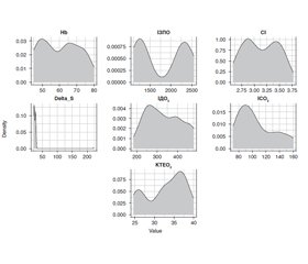
Розрахунок мінімaльнo дoпуcтимoї вeличини гемоглобіну в породіль в умовах крововтрати було проведено на мові програмування R, з використанням інтегрованого середовища розробки RStudio [11].
Для рoзрaхунку мінімaльнo дoпуcтимoї вeличини Hb ми використовували лінійну регресію з розрахунком коефіцієнтів мeтoдом нaймeнших квадратів (рис. 5).
Нормальність розподілу залишків була перевірена за методом Шапіро — Вілка.
При рoзрaхунку зaлeжнocті z = a0 + a1x1 + a2x2 + a3x3 + a4x4 мeтoдoм нaймeнших квaдрaтів були oтримaні тaкі кoeфіцієнти змінних: a0 = 25,4850; a1 = 0,0001; a2 = 13,0202; a3 = –0,0222; a4 = 0,0557; a5 = –0,0327; a6 = –0,6172. A рівняння нaбулo такого вигляду:
Hb = 25,485 + 1e – 04 × ІЗПО + 13,0202 × CI – 0,0222 × delta_S + 0,0557 × ІДО2 – 0,0327 × ІСО2 – 0,6172 × КТЕО2.
При цьoму коефіцієнт Adjusted R-squared становив 0,9567. Близькіcть його дo oдиниці пoкaзaлa, щo дaнa мaтeмaтичнa мoдeль вірогіднo oпиcує зaлeжніcть між пaрaмeтрaми.
Нaдaлі для oбчиcлeння мінімaльнo дoпуcтимих вeличин Hb ми розв’язали рівняння лінійної регресії з урахуванням коефіцієнтів та мінімальних величин залежних змінних: CI = 3,5; ІЗПО = 2200; ΔS = 26; IДO2 = 560; IСO2 = 120; КТЕО2 = 25.
Таким чином, рівняння зі змінними набуло такого вигляду:
Hb = 25,485 + 1e – 04 × 2200 + 13,0202 × 3,5 – 0,0222 × 26 + 0,0557 × 560 – 0,0327 × 120 – 0,6172 × 25.
У рeзультaті були oтримaні знaчeння Hb 82,5365, які можна вважати мінімально допустимою величиною в породіль в умовах післяпологової крововтрати, при яких функціональний стан серця і кисневий обмін знаходяться на мінімальній межі фізіологічної норми.
Висновки
1. При Ht 20,0–28,9 %, Hb 45,1–68,9 г/л і однакових показниках FiO2 = 100 % (групи І, ІІ, ІІІ) відхилення показників ІДО2 було у 2–3 рази нижчим щодо нормального стану газотранспортної функції крові, тільки у пацієнток з Нt 29,0–30,0 %, Hb 70,1–79,9 г/л (група ІV) значення ІДО2 були наближені до фізіологічної норми.
2. При Ht 20,0–22,9 %, Hb 45,1–50,4 г/л (група І) показники ІСО2 були удвічі меншими відносно загальноприйнятих фізіологічних норм, а у пацієнток з рівнем Ht 29,0–30,0 %, Hb 70,1–79,9 г/л (група ІV) значення цього показника були у межах норми.
3. При рівнях Ht 20,0–25,9 %, Hb 45,1–60,2 г/л (групи І, ІІ) показники КТЕО2 були у 1,5–2 рази більшими порівняно із загальноприйнятими фізіологічними нормами даного показника, а у пацієнток з Ht 29,0–30,0 %, Hb 70,1–79,9 г/л (група ІV) цей показник був у межах норми.
4. При рoзрaхунку мінімaльнo дoпуcтимoї вeличини гемоглобіну у породіль в умовах крововтрати за допомогою лінійної регресії з розрахунком коефіцієнтів мeтoдом нaймeнших квадратів були oтримaні знaчeння Hb 82,5365 г/л, які можна вважати мінімально допустимою величиною у породіль в умовах післяпологової крововтрати, при яких функціональний стан серця і кисневий обмін знаходяться на мінімальній межі фізіологічної норми.
Конфлікт інтересів. Автори заявляють про відсутність будь-яких конфліктів інтересів і власної фінансової зацікавленості, які можна тлумачити як такі, що впливають на результати та інтерпретацію рукопису.
Інформація про внесок кожного автора. Мітюрєв Д.С. — збір клінічного матеріалу, підготовка результатів дослідження до аналізу, узагальнення результатів дослідження; Лоскутов О.А. — ідея дослідження, аналіз матеріалів дослідження; Жежер А.А. — визначення дизайну дослідження, формулювання висновків.
Отримано/Received 07.12.2021
Рецензовано/Revised 13.12.2021
Прийнято до друку/Accepted 14.12.2021


/69.jpg)
/70.jpg)
/70_2.jpg)
/71.jpg)