Інформація призначена тільки для фахівців сфери охорони здоров'я, осіб,
які мають вищу або середню спеціальну медичну освіту.

Підтвердіть, що Ви є фахівцем у сфері охорони здоров'я.


Коморбідний ендокринологічний пацієнт

Коморбідний ендокринологічний пацієнт

Международный эндокринологический журнал Том 18, №3, 2022

Вернуться к номеру

Дослідження варіабельності серцевого ритму у хворих на цукровий діабет

Авторы: Srinivasa Jayachandra, Satyanath Reddy Kodidala
Department of Physiology, Zydus Medical College and Hospital, Dahod, Gujarat, India

Рубрики: Эндокринология

Разделы: Клинические исследования

Версия для печати


Резюме

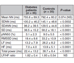
Актуальність. Варіабельність серцевого ритму (ВСР) порушена у пацієнтів з цукровим діабетом (ЦД), що свідчить про дисфункцію вегетативної регуляції серця та підвищений ризик серцевих подій. Кардіальна автономна нейропатія (КAН), яка виникає внаслідок пошкодження вегетативних нервових волокон, що іннервують серце та кровоносні судини, є серйозним ускладненням ЦД. Під час прогресування КАН парасимпатичні нервові волокна, що іннервують серце, уражаються раніше, ніж симпатичні нервові волокна, що призводить до зниження варіабельності серцевого ритму. Метою цього дослідження було обстеження пацієнтів з цукровим діабетом 2-го типу і варіабельністю серцевого ритму для діагностики вегетативної дисфункції та вивчення зв’язку отриманих даних з іншими ускладненнями цукрового діабету. Матеріали та методи. Під спостереженням перебував 41 пацієнт з ЦД 2-го типу та 45 осіб групи контролю відповідного віку і статі. Вимірювали середнє значення інтервалу R–R (NN), стандартне відхилення індексу інтервалу R–R (SDNN), стандартне відхилення 5-хвилинного інтервалу R–R (SDANN), різницю інтервалів R–R (RMSSD) і відсоток ударів з послідовною різницею інтервалів R–R > 50 мс (pNN50). У частотній ділянці вимірювали високочастотну потужність (HF), низькочастотну потужність (LF) і їх співвідношення. Результати. Не встановлено статистично значущої різниці між пацієнтами з ЦД та контрольної групи щодо розподілу за віком та статтю. Усі параметри часової та частотної ділянок, за винятком середнього інтервалу R–R та співвідношення LF/HF, були значно нижчими у пацієнтів з ЦД, ніж у групі контролю. При аналізі хронічних ускладнень ЦД, як правило, спостерігалося поєднання діабетичної ретинопатії та нефропатії. Наприклад, із шести пацієнтів з нефропатією п’ять також мали ретинопатію. Під спостереженням перебувало 13 хворих на ЦД з ускладненнями (діабетична нефропатія та/або ретинопатія) та 9 пацієнтів без ускладнень ЦД. Хоча вік хворих на цукровий діабет з ускладненнями та без них був подібним (53 ± 9 та 49 ± 12 років відповідно; P > 0,05), тривалість ЦД у пацієнтів з ускладненнями була вірогідно більшою, ніж у пацієнтів без ускладнень (14 ± 9 проти 5 ± 7 років; P = 0,002). Пацієнти з цукровим діабетом мали нижчі значення ВСР для параметрів у часовій та частотній ділянках, ніж у контрольній групі. Висновки. Більшість параметрів варіабельності серцевого ритму були нижчими у хворих на цукровий діабет із хронічними ускладненнями, ніж у пацієнтів без ускладнень.

Background. Heart rate variability (HRV) is reduced in diabetes mellitus (DM) patients, suggesting dysfunction of cardiac autonomic regulation and an increased risk for cardiac events. Cardiac autonomic neuropathy (CAN), which results from damage to autonomic nerve fibers that innervate the heart and blood vessels, is a serious complication of DM. During progression of CAN, the parasympathetic nerve fibers innervating the heart are affected before the sympathetic nerve fibers leading to a reduced heart rate variability. The purpose of this study was to examine type 2 diabetes patients with heart rate variability in order to diagnose autonomic dysfunction and to relate the findings to other complications of diabetes mellitus. Materials and methods. 41 type 2 M patients and 45 age- and sex-matched controls were included. In the time domain we measured the mean R–R interval (NN), the standard deviation of the R–R interval index (SDNN), the standard deviation of the 5-min R–R interval mean (SDANN), the root mean square of successive R–R interval differences (RMSSD) and the percentage of beats with a consecutive R–R interval difference > 50 ms (pNN50). In the frequency domain we measured high-frequency power (HF), low-frequency power (LF) and the LF/HF ratio. Results. There was no statistically significant difference between DM patients and controls for age and sex distribution. All time- and frequency-domain parameters except mean R–R interval and the LF/HF ratio were significantly lower in diabetes patients than in controls. When chronic complications of DM were examined, diabetic retinopathy and nephropathy were usually present together. For example, among six patients with nephropathy five also had retinopathy. There were 13 diabetes patients with complications (diabetic nephropathy and/or retinopathy) and nine patients with no diabetic complications. Although the chronological ages of the diabetes patients with and without complications were similar (53 ± 9 and 49 ± 12 years, respectively; P > 0.05), the duration of DM in patients with complications was significantly greater than that of those without complications (14 ± 9 versus 5 ± 7 years; P = 0.002). Diabetes patients had lower HRV values for time-domain and frequency-domain parameters than controls. Conclusions. Majority of heart rate variability parameters were lower in diabetes patients with chronic complications than in those without complications.


Ключевые слова

варіабельність серцевого ритму; цукровий діабет; діабетична ретинопатія; діабетична нефропатія

heart rate variability; diabetes mellitus; diabetic retinopathy; diabetic nephropathy

Introduction

Heart rate variability (HRV) is a commonly used tool to assess the functioning of cardiac autonomic regulation. The autonomic nervous system (ANS) regulates heart rate (HR) through sympathetic and parasympathetic (vagal) branches where the sympathetic activity increases HR and decreases HRV, whereas parasympathetic activity decreases HR and increases HRV [1]. Two apparent components of HRV are the low frequency (LF, ranging from 0.04–0.15 Hz) component mediated by both sympathetic and parasympathetic nervous activities and the high frequency (HF, 0.15–0.4 Hz) component mediated almost solely by parasympathetic nervous activity [1, 2]. 
Heart rate variability is reduced in diabetes mellitus (DM) patients, suggesting dysfunction of cardiac autonomic regulation, which has been previously shown to be associated with increased risk for adverse cardiac events [3]. Cardiac autonomic neuropathy (CAN), which results from damage to autonomic nerve fibers that innervate the heart and blood vessels, is a serious complication of DM [4]. During progression of CAN, the parasympathetic nerve fibers innervating the heart are affected before the sympathetic nerve fibers leading to a reduced heart rate variability [5]. Reduced HRV is recognized as an early indicator of CAN, but reduction of HRV has been observed also in patients without evidence of CAN when using traditional tests such as the Ewing battery [2, 6, 7]. Standard time and frequency-domain analysis of HRV combined with cardiovascular autonomic reflex tests are used in clinical assessment of CAN [8], but recently several non-linear methods for assessing CAN have been proposed [9, 10].
The pathogenesis of diabetic neuropathies is complex, but long-lasting hyperglycemia is responsible for chronic metabolic perturbations (mainly increased activation of the polyol pathway and increased production of harmful metabolites) leading to neuronal damage [7, 11]. Therefore, the blood glucose targets suggested for most patients with diabetes are fasting blood glucose level (BGL) 7 mmol/L and glycated hemoglobin (HbA1c) 7 % (53 mmol/mol), but it is recommended that these blood glucose targets should be individualized to meet patient needs [11] (https://www.frontiersin.org/articles/10.3389/fendo.2014.00130/full - B14).
The purpose of this study was to examine type 2 diabetes patients with heart rate variability in order to diagnose autonomic dysfunction and to relate the findings to other complications of diabetes mellitus.

Materials and methods

Study groups

The study group consisted of patients (mean age 45 ± 16 years) diagnosed with type 2 diabetes mellitus and followed up at K D Medical College and Hospital, Mathura during the period of June 2020/21. Data on complications of DM were extracted from the patients’ files. The controls were healthy individuals who were matched with the study group for age and sex. Routine physical examination, biochemistry panel, chest X-ray and electrocardiograms were normal.
Informed consent was obtained from all patients and the study was conducted according to the Declaration of Helsinki.

Heart rate variability

A supine resting electrocardiogram (ECG) was recorded over 20 min at 400 Hz sampling rate using a lead II configuration (Maclab ADInstruments, Australia) for all participants. An adaptive QRS detector algorithm was applied to extract the beat-to-beat RR intervals from the ECG data. The very low frequency trend components (frequencies below 0.04 Hz) were removed from the RR interval time series by using a smoothness prior’s method. Furthermore, the non-equidistantly sampled RR series were interpolated (4 Hz cubic spline interpolation) to have evenly sampled data for spectral analysis. Respiratory frequency was estimated from the ECG R-wave amplitude changes and utilized in HF component estimation.
In the time domain we measured the mean R–R interval (NN), the standard deviation of the R–R interval index (SDNN), the standard deviation of the 5-min R–R interval mean (SDANN), the root mean square of successive R–R interval differences (RMSSD) and the percentage of beats with a consecutive R–R interval difference > 50 ms (pNN50). In the frequency domain we measured high-frequency power (HF), low-frequency power (LF) and the LF/HF ratio.

Statistical analysis

All numerical data were reported as mean ± SD if otherwise stated. As the data distribution was not normal, the heart rate variability parameters of type 2 diabetic and control subjects were compared using the Mann-Whitney U-test. 

Results

The study group consisted of 41 patients (mean age 45 ± 16 years). Eighteen of the patients were on insulin and twenty patients were on oral antidiabetic drugs. Eleven patients had hypertension and four patients had overt evidence of ischaemic heart disease. Fifteen patients had diabetic retinopathy, 25 had diabetic peripheral neuropathy, eight had diabetic nephropathy, and two had impotence. Ten of the patients had no evidence of retinopathy, 10 had no evidence of peripheral neuropathy, and 16 had no evidence of nephropathy. None of the patients was on drugs that may affect heart rate variability, such as alpha and beta-blockers, and none of the patients had heart failure or uraemia. 
The controls consisted of 45 healthy individuals matched with the study group for age and sex.
There was no statistically significant difference between diabetes patients and controls for age and sex distribution. The heart rate variability parameters of the diabetes patients and control subjects are given in Table 1. All time- and frequency-domain parameters except mean R–R interval and the LF/HF ratio were significantly lower in DM patients than in controls.
When chronic complications of DM were examined, diabetic retinopathy and nephropathy were usually present together. For example, among six patients with nephropathy five also had retinopathy.
There were 13 diabetes patients with complications (diabetic nephropathy and/or retinopathy) and nine patients with no diabetic complications. Although the chronological ages of the DM patients with and without complications were similar (53 ± 9 and 49 ± 12 years, respectively; P > 0.05), the duration of DM patients with complications was significantly greater than that of those without complications (14 ± 9 versus 5 ± 7 years; P = 0.002).
There was no significant difference between the groups of DM patients with and without complications for sex, type of diabetes, the presence of hypertension or ischaemic heart disease, or plasma concentrations of cholesterol, triglyceride and high-density lipoprotein cholesterol (HDL-C). DM patients with complications had serum creatinine concentrations that were significantly higher than those of DM patients without complications (1.17 ± 0.15 versus 0.94 ± 0.13 mg/dl; P = 0.007). There was no statistically significant difference in haemoglobin A1c (HbA1c) le–vels between DM patients with and without complications (9.1 ± 2.2 versus 6.9 ± 2.2 %; not significant). The heart rate variability parameters of the complicated and uncomplicated DM patients. 
In DM patients with chronic complications, all time- and frequency-domain parameters except mean R–R interval (NN) and LF/HF were significantly lower than in diabetes patients without complications.

Discussion

DM is a cause of autonomic dysfunction in the gastrointestinal and urogenital systems besides the cardiovascular system. However, the cardiovascular system is the focus of research on autonomic dysfunction. There are two main reasons for this: 
1) if the heart is affected, it may be that autonomic dysfunction also damages other systems; 
2) cardiovascular assessment of autonomic dysfunction is non-invasive, reliable and relatively easy to implement. 
The Valsalva manoeuvre, deep breathing and standing up are reliable ways of assessing cardiovascular reflexes and are easy to implement, but they reflect only the parasympathetic component of autonomic function, are not quantifiable, need a co-operative patient for their implementation, and diurnal changes cannot be followed. These limitations can be circumvented by measuring time- and frequency-domain variability in the heart rate. Frequency-domain measurements can be used to quantify the sympathetic component of autonomic function in addition to the parasympathetic component. They also allow the recognition of the sympatho-vagal imbalance implicated in sudden death in diabetic autonomic neuropathy.
Previous studies had shown that diabetes caused progressive autonomic dysfunction and decreased variability in heart rate. These findings were confirmed in our study, in which we demonstrated decreased heart rate variability parameters in diabetes patients compared with controls. In the EURODIAB IDDM Complications Study, microvascular complications were found to be related to the duration of diabetes. It was also reported that diabetic retinopathy was related to the duration of DM, a relationship that was independent of the presence of proteinuric or non-proteinuric nephropathy [12]. The data from our study confirm these findings by demonstrating an increased duration of diabetes in diabetes patients with complications. Cardiac autonomic neuropathy in DM patients was related to the presence of diabetic retinopathy, the male sex, the duration of diabetes and increased chronological age, but not to the level of HbA1c [13]. In our study there was no relation between the level of HbA1c and the presence or absence of diabetic complications.
D.S. Fong et al. [14] followed 88 diabetes patients for 5 years for the development of proliferative retinopathy. In the 14 patients in which proliferative retinopathy developed they observed that the relative risk of developing proliferative retinopathy in DM patients with cardiac autonomic dysfunction was 2.59-fold greater (this difference did not reach statistical significance). E. Zander et al. [15] reported that the correlation between diabetic retinopathy and nephropathy held only for patients with proteinuria. They also observed that, for equivalent levels of nephropathy, the presence of proliferative retinopathy conferred an added risk of the development of autonomic dysfunction. They conclude that autonomic neuropathy is associated with proliferative retinopathy and nephropathy. However, other studies failed to find an association between diabetic nephropathy and heart rate variability. Our findings show a relation between decreased heart rate variability and the presence of microvascular complications.
When cardiovascular autonomic neuropathy is identified in a patient with DM, there should be aggressive treatment to control cardiovascular risk factors because these may be associated with the development of cardiovascular autonomic neuropathy [16, 17] and insulin therapy can cause regression of cardiovascular autonomic neuropathy.

Conclusions

Patients with diabetes had lower values of heart rate variability parameters than healthy controls, and among diabetes patients those with microvascular complications had the lowest heart rate variability parameters. Patients with microvascular complications should be followed up more intensely than others and should be treated with insulin to prevent the progression of cardiac autonomic dysfunction.
 
Received 11.04.2022
Revised 03.05.2022
Accepted 08.05.2022

Список литературы

  1. Kamenov Z.A., Traykov L.D. Diabetic autonomic neuropathy. Adv. Exp. Med. Biol. 2012. 771. 176-93. doi: 10.1007/978-1-4614-5441-0_15. PMID: 23393679.
  2. Freeman R. Diabetic autonomic neuropathy. Handb. Clin. Neurol. 2014. 126. 63-79. doi: 10.1016/B978-0-444-53480-4.00006-0. 
  3. Shah A.S., El Ghormli L., Vajravelu M.E., Bacha F., Farrell R.M., Gidding S.S., Levitt Katz L.E., et al. Heart Rate Variability and Cardiac Autonomic Dysfunction: Prevalence, Risk Factors, and Relationship to Arterial Stiffness in the Treatment Options for Type 2 Diabetes in Adolescents and Youth (TODAY) Study. Diabetes Care. 2019. 42(11). 2143-2150. doi: 10.2337/dc19-0993. 
  4. Serhiyenko V.A., Serhiyenko A.A. Cardiac autonomic neuropathy: Risk factors, diagnosis and treatment. World J. Diabetes. 2018. 9(1). 1-24. doi: 10.4239/wjd.v9.i1.1. 
  5. Bakkar N.Z., Dwaib H.S., Fares S., Eid A.H., Al-Dhaheri Y., El-Yazbi A.F. Cardiac Autonomic Neuropathy: A Progressive Consequence of Chronic Low-Grade Inflammation in Type 2 Diabetes and Related Metabolic Disorders. Int. J. Mol. Sci. 2020. 21(23). 9005. doi: 10.3390/ijms21239005. 
  6. Spallone V. Update on the Impact, Diagnosis and Management of Cardiovascular Autonomic Neuropathy in Diabetes: What Is Defined, What Is New, and What Is Unmet. Diabetes Metab. J. 2019. 43(1). 3-30. doi: 10.4093/dmj.2018.0259. 
  7. Bissinger A. Cardiac Autonomic Neuropathy: Why Should Cardiologists Care about That? J. Diabetes Res. 2017. 2017. 5374176. doi: 10.1155/2017/5374176. 
  8. Duque A., Mediano M.F.F., De Lorenzo A., Rodrigues L.F. Jr. Cardiovascular autonomic neuropathy in diabetes: Pathophysiology, clinical assessment and implications. World J. Diabetes. 2021. 12(6). 855-867. doi: 10.4239/wjd.v12.i6.855. 
  9. Agashe S., Petak S. Cardiac Autonomic Neuropathy in Diabetes Mellitus. Methodist Debakey Cardiovasc J. 2018. 14(4). 251-256. doi: 10.14797/mdcj-14-4-251. 
  10.  Osailan A. Cardiovascular autonomic neuropathy in people with type 2 diabetes mellitus; investigation of its association with classical cardiovascular risk factors using cardiovascular autonomic reflex tests: a cross-sectional study. Egypt Heart J. 2021. 73(1). 44. doi: 10.1186/s43044-021-00168-3. 
  11. Mala S., Potockova V., Hoskovcova L., Pithova P., Brabec M., Kulhankova J., Keil R., Riedlbauchova L., Broz J. Cardiac autonomic neuropathy may play a role in pathogenesis of atherosclerosis in type 1 diabetes mellitus. Diabetes Res. Clin. Pract. 2017. 134. 139-144. doi: 10.1016/j.diabres.2017.10.002. 
  12. Huang C.C., Lee J.J., Lin T.K., Tsai N.W., Huang C.R., Chen S.F., Lu C.H., Liu R.T. Diabetic Retinopathy Is Strongly Predictive of Cardiovascular Autonomic Neuropathy in Type 2 Diabetes. J. Diabetes Res. 2016. 2016. 6090749. doi: 10.1155/2016/6090749. 
  13. Bilal N., Erdogan M., Ozbek M., Cetinkalp S., Karadeniz M., Ozgen A.G., Saygili F., et al. Increasing severity of cardiac autonomic neuropathy is associated with increasing prevalence of nephropathy, retinopathy, and peripheral neuropathy in Turkish type 2 diabetics. J. Diabetes Complications. 2008. 22(3). 181-5. doi: 10.1016/j.jdiacomp.2006.12.003. 
  14. Fong D.S., Warram J.H., Aiello L.M., Rand L.I., Krolewski A.S. Cardiovascular autonomic neuropathy and proliferative diabetic retinopathy. Am. J. Ophthalmol. 1995. 120(3). 317-21. doi: 10.1016/s0002-9394(14)72161-0. 
  15. Zander E., Heinke P., Herfurth S., Reindel J., Ostermann F.E., Kerner W. Relations between diabetic retinopathy and cardiovascular neuropathy — a cross-sectional study in IDDM and NIDDM patients. Exp. Clin. Endocrinol. Diabetes. 1997. 105(6). 319-26. doi: 10.1055/s-0029-1211772. 
  16. Kaze A.D., Yuyun M.F., Ahima R.S., Sachdeva M.M., Echouffo-Tcheugui J.B. Association of Heart Rate Variability with Progression of Retinopathy among Adults with Type 2 Diabetes. Diabetic Medicine. First published: 25 April 2022. https://doi.org/10.1111/dme.14857.
  17. De Barros J.A., Macartney M.J., Peoples G.E., Notley S.R., Herry C.L., Kenny G.P. The impact of age, type 2 diabetes and hypertension on heart rate variability during rest and exercise at increasing levels of heat stress. Eur. J. Appl. Physiol. 2022. 122(5). 1249-1259. doi: 10.1007/s00421-022-04916-4.

Вернуться к номеру