Інформація призначена тільки для фахівців сфери охорони здоров'я, осіб,
які мають вищу або середню спеціальну медичну освіту.

Підтвердіть, що Ви є фахівцем у сфері охорони здоров'я.


Коморбідний ендокринологічний пацієнт

Коморбідний ендокринологічний пацієнт

Международный эндокринологический журнал Том 18, №4, 2022

Вернуться к номеру

The role of vitamin D for the management of depression in patients with autoimmune thyroiditis and hypothyroidism in the West-Ukrainian population

Авторы: Iryna Kamyshna
I. Horbachevsky Ternopil National Medical University, Ternopil, Ukraine

Рубрики: Эндокринология

Разделы: Клинические исследования

Версия для печати


Резюме

Актуальність. Відомо, що тиреоїдит Хашимото є одним із найчастіших ендокринних захворювань, яке вражає населення різних вікових груп і може призвести до гіпотиреозу. Ця патологія також є одним із найбільш поширених авто­імунних захворювань. Пацієнти з гіпотиреозом часто відчувають ознаки депресії, яка переважає серед інших симптомів гіпотиреозу. Дані останніх досліджень довели, що дефіцит вітаміну D може викликати прояви депресії в населення. Метою проведеної роботи є вивчення впливу холекальциферолу в пацієнтів з автоімунним тиреоїдитом та гіпотиреозом у західноукраїнській популяції на рівень депресії у цих хворих. Матеріали та методи. У дослідження були включені 56 пацієнтів із гіпотиреозом, спричиненим автоімунним тиреоїдитом. Визначали наявність депресії за допомогою шкали депресії Гамільтона (HDRS). Обстеження проводили на початку та в кінці 12-тижневого лікування. Результати. У па­цієнтів першої групи, які отримували холекальциферол і L-тироксин, рівень депресії за шкалою Гамільтона знизився на 40 %, тоді як у пацієнтів, які отримували тільки L-тироксин, рівень депресії знизився на 25 %. Крім того, спостерігалася вірогідна різниця між пацієнтами першої та другої груп після лікування (р = 0,003). При аналізі ефекту лікування в пацієнтів першої групи з додатковим призначенням холекальциферолу на тлі L-тироксину у 21,4 % пацієнтів зникли прояви депресії. Крім того, спостерігалося зниження рівня депресії у решти пацієнтів цієї групи. Ступінь вираженості депресивних проявів зменшився від середньої тяжкості до легкого депресивного розладу. Висновки. Пацієнтам, які страждають на автоімунний тиреоїдит та гіпотиреоз, слід призначати вітамін D, що сприяє зменшенню в них частоти депресивних розладів.

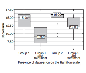
Background. Hashimoto’s thyroiditis is known to be an essential endocrine disease that affects the population and may lead to hypothyroidism. This disease is one of the most commonly spread autoimmune di­seases. Hypothyroid patients frequently experience features of depression, which is prevalent among other symptoms in hypothyroidism. Data from recent research has proved that vitamin D deficiency may cause depression manifestations in the population. The purpose of the work is to study the effect of cholecalciferol in patients with autoimmune thyroiditis and hypothyroidism in the West-Ukrainian population on the level of depression in these patients. Materials and methods. The study included the 56 patients with hypothyroidism (H) caused by autoimmune thyroiditis (AIT). We identified the severity of depression levels using the Hamilton Depression Rating Scale (HDRS), which is reliable for depression assessment. Examinations were performed at the beginning and end of the 12-week treatment. Results. In patients of group 1 who received cholecalciferol and L-thyroxine, the level of depression on the Hamilton scale decreased by 40 %, while in patients who received only L-thyroxine, the level of depression decreased by 25 %. In addition, there was a significant difference between patients in groups 1 and 2 after treatment (p = 0.003). That is, treatment with additional cholecalciferol on the background of L-thyroxine was more effective than treatment with L-thyroxine alone. Analyzing the effect of treatment in patients with Group 1 with the additional appointment of cholecalciferol on the background of L-thyroxine in 21.4 % of patients disappeared depression. In addition, there was a decrease in depression in other patients in this group. Thus, in the remaining patients the severity of depressive manifestations decreased from moderate severity to mild depressive disorder. At the same time, after treatment only L-thyroxine depressive disorder of moderate severity decreased from 78.6 to 35.7 % to mild depressive disorder, but complete disappearance of depression in this group of patients after treatment was not observed. Conclusions. Vitamin D supplementation should be administered in patients suffering from autoimmune thyroiditis and hypothyroidism which may correct depression disorders in these patients.


Ключевые слова

автоімунний тиреоїдит; гіпотиреоз; депресія

autoimmune thyroiditis; hypothyroidism; depression

Introduction

Hashimoto’s thyroiditis (HT) is known to be an essential endocrine disease that affects the population and may lead to hypothyroidism. This disease is one of the most commonly spread autoimmune diseases [1]. Hypothyroid patients frequently experience features of depression which is prevalent among other symptoms in hypothyroidism [2]. In terms of thyroid disorders, the rate of depression moderately increases in patients suffering from hypothyroidism [3]. Different polymorphisms of vitamin D receptors highly contribute to the risk development for autoimmune thyroiditis (AIT) [4]. Data from recent research has proved that vitamin D deficiency may cause depression manifestations in the population generally [5]. 
The insufficient intake of vitamin D is a common problem throughout the world, and people lacking vitamin D are more likely at the risk of experiencing major depressive disorder. Therefore, vitamin D supplementation is endorsed for the prevention and treatment of such conditions. Although preclinical investigations demonstrate limited and poor-quality evidence on the feasible mechanisms based on the favorable effects of vitamin D for the therapy of these disorders, most of the clinical studies prove that vitamin D supplementation enhances the reduction of the prevalence of depressive symptoms [6]. 
The purpose of our work is to study the effect of cholecalciferol in patients with autoimmune thyroiditis and hypothyroidism in the West-Ukrainian population on the level of depression in these patients.

Materials and methods

Our research was conducted in Bukovinian State Medical University, Chernivtsi Regional Endocrinology Center, and I. Horbachevsky Ternopil National Medical University, Ukraine. 
The study included the 56 patients with hypothyroidism (H) caused by AIT. These patients were distributed into two groups. Patients in the first group (n = 28) received cholecalciferol at a dose of 4000 IU/day (28,000 IU/week) and L-thyroxine (88.39 ± 12.70 μg/day). Patients in the second group (n = 28) were prescribed only L-thyroxine (87.50 ± 12.73 μg/day). Examinations were performed at the beginning and end of the 12-week treatment.
Ethical approval. The study fully ensured standards described in the 1975 Helsinki Declaration of Human Rights (amended in 2008). The participants completed and signed a written informed consent before enrolling voluntarily in the research. Ethical approvation was taken from I. Horbachevsky Ternopil National Medical University (09.02.2021).
To diagnose hypothyroidism, we were guided by recommendations required by the American Association of Clinical Endocrinologists 2012 [7]. The corresponding clinical features were considered when verifying AIT, namely the results of a sonogram of the thyroid gland (reduced echogenicity) and circulating antibodies antibodies against thyroid peroxidase (TPO-Ab), and thyroglobulin (TG-Ab) were detected.
Blood samples from patients and controls were taken in the morning (8 to 10 am) after a night fast. Using STAT FAX303/Plus analyzer (Awareness Technology Inc, USA), we determined levels of free thyroxine (fT4, normal range 6.0–13.0 pmol/L for males and 7.0–13.5 pmol/L for females), thyroid-stimulating hormone (TSH, normal range 0.3–4.0 mIU/mL), TPO-Ab (normal range 0–30 IU/mL) and TG-Ab (normal range 0–65 IU/mL) in each individual who participated in the study.
Study exclusion criteria were the following: less than 18 years of age, malignancy, inflammation resulting from rheumatic diseases or acute/chronic infection, diabetes mellitus, vascular, chronic diseases of liver and kidneys, and pregnancy. Individuals administering drugs that could influence thyroid function were also ruled out from the study.
We identified the severity of depression levels using the Hamilton Depression Rating Scale (HDRS), which is reliable for depression assessment. Due to the HDRS, scores of 0–7, 8–13, 14–18, 19–23, and 23–53 are regarded as normal, mild, medium, severe, and very severe, correspondingly [8].
Statistical analysis. Quantitative variables were assessed for normality using the Shapiro-Wilk test (when the number of subjects was less than 50) or the Kolmogorov-Smirnov test (when the number of subjects was more than 50). Quantitative variables following non normal distribution were described using median (Me) and lower and upper quartiles (Q1–Q3). Comparisons of three or more groups on a quantitative variable whose distribution differed from normal were made using the Kruskal-Wallis test and Dunn’s criterion with Holm correction as a post-hoc method. Comparison of frequencies in the analysis of multifield contingency tables was performed using Pearson’s chi-square test (for expected values greater than 10).

Results 

In our study, after treatment, there was a decrease in TPO-Ab levels in patients taking cholecalciferol and L-thyroxine by 31.25 % (p < 0.001), while in patients in group 2 who received only L-thyroxine, TPO-Ab levels decreased only at 14.1 % (p < 0.05)
When comparing TG-Ab after treatment, there was a significant decrease in TG-Ab levels in patients receiving cholecalciferol and L-thyroxine by 18.84 % (p < 0.001), while in patients of group 2 who received only L-thyroxine TPO-Ab level decreased by only 8.82 %.
We performed analysis of depression conditioning on presence of depression on the Hamilton scale.
After treatment, there is a decrease in the level of depression on the Hamilton scale (Table 2). Thus, in patients of group 1 who received cholecalciferol and L-thyroxine, the level of depression on the Hamilton scale decreased by 40 %, while in patients who received only L-thyroxine, the level of depression decreased by 25 %. In addition, there was a significant difference between patients in groups 1 and 2 after treatment (p = 0.003). That is, treatment with additional cholecalciferol on the background of L-thyroxine was more effective than treatment with L-thyroxine alone.
Analyzing the effect of treatment in patients with Group 1 with the additional appointment of cholecalciferol on the background of L-thyroxine in 21.4 % of patients depression disappeared. In addition, there was a decrease in depression frequency in other patients in this group (Figure 1). Thus, in the remaining patients the severity of depressive manifestations decreased from moderate severity to mild depressive disorder.
At the same time, after treatment only L-thyroxine depressive disorder of moderate severity decreased from 78.6 to 35.7 % to mild depressive disorder, but complete disappearance of depression in this group of patients after treatment was not observed (Figure 2).

Discussion

Thyroid dysfunctions are closely connected with deterioration of brain function [9]. The valuable insights on the correlation between thyroid hormone deficiency with depression have been discussed substantially in various credible studies [10]. Symptoms of depression tend to coincide in patients. However, the treatment of these disorders remains controversial. Vitamin D deficiency is a universal health problem. The prevalence of vitamin D deficiency or insufficiency exceeds a billion worldwide [11]. 
HT is considered chronic autoimmune thyroiditis characterized by lymphocytic infiltration, which may finally lead to the progressive loss of thyroid tissue [12]. Some studies prove that HT is the outcome of the united action of genetic susceptibility and environmental factors, but the precise route is still not established [13]. One study claimed that in the case of normal thyroid function, HT could also induce neuro-inflammation, resulting in emotional alterations [14]. Nowadays, the possibilities of HT are also gro–wing; most subjects do not manifest symptoms, which are commonly observed at the physical checkups. Some studies have favored that vitamin D produces a positive protective influence on autoimmune thyroid diseases and thyroid malignancies, but the mechanism has not been elucidated [15].
One essential of the numerous functions of vitamin D implies the regulation of nervous system development in addition to being involved in calcium regulation [16]. Vitamin D attenuates and easily passes the blood-brain barrier [17]. Vitamin D receptors are widely found in different brain sites, i.e., the prefrontal cortex, substantia nigra, and hypothalamus [18]. Vitamin D is also closely connected with the synthesis of monoamines, such as serotonin, dopamine, and norepinephrine, and its steroid properties enable modulating the activity of GABA-A receptors [18–20]. Furthermore, several studies have suggested that vitamin D is related to nerve growth factor enhancement and antioxidant effects in the central nervous system [20–22]. 
A meta-analysis identified the impact of vitamin D administration on depression. The findings reported positive results of vitamin D for the treatment of depression [23].
In our study, after treatment with additional administration of cholecalciferol on the background of L-thyroxine in 21.4 % of patients disappeared depression. In addition, there was a reduction in depression in other patients in this group. Thus, in the remaining patients the severity of depressive symptoms decreased from moderate to mild depressive disorder.
For the last three decades, a complicated association between peripheral immune activation, neuroinflammation, and changes in brain circuits linked to depression has been traced [24]. In this respect, vitamin D is considered a well-known regulator of innate immunity that acts not only as transcription and a growth factor but also interacts with surface receptors in different immune cells [25]. Therefore, numerous positive effects of vitamin D influencing the behavior might be related to its capability to stabilize peripheral and central nervous system (CNS) immune responses.
In the CNS, 1.25(OH)D is often connected with neuroprotective properties. A contemporary study proved that 1,25(OH)D was able to inhibit serotonin reuptake transport and monoamine oxidase-A gene expression (MAO-A; the enzyme which is in charge of monoamine degradation) in cultured rat serotonergic neuronal cell lines, presupposing that calcitriol tends to act similarly to antidepressants [26]. 
In our study after treatment, there is a decrease in the level of depression on the Hamilton scale. Thus, in patients of Group 1 who received cholecalciferol and L-thyroxine, the level of depression on the Hamilton scale decreased by 40 %, while in patients who received only L-thyroxine, the level of depression decreased by 25 %. That is, treatment with additional cholecalciferol on the background of L-thyroxine was more effective than treatment with L-thyroxine alone. Regarding that vitamin D contributes to neuro-immunomodulation and neuroplasticity and may decrease oxidative stress [27], it has been researched as a possible strategy for the treatment of symptoms of depression.

Conclusions

Considering our results, vitamin D3 supplementation should be administered in patients suffering from autoimmune thyroiditis and hypothyroidism which may correct depression disorders in these patients.
Ongoing and prospective long-term randomized controlled trials are necessary to gain insight into the effectiveness and safety of Vitamin D as a therapeutic agent for these thyroid disorders and the prevention and treatment of neurological complications.
 
Received 18.05.2022
Revised 11.06.2022
Accepted 15.06.2022

Список литературы

  1. Caturegli P., De Remigis A., Rose N.R. Hashimoto thyroiditis: clinical and diagnostic criteria. Autoimmun. Rev. 2014. 13(4-5). 391-7. doi: 10.1016/j.autrev.2014.01.007. 
  2. Gorkhali B., Sharma S., Amatya M., Acharya D., Sharma M. Anxiety and Depression among Patients with Thyroid Function Disorders. J. Nepal Health Res. Counc. 2020. 18(3). 373-378. doi: 10.33314/jnhrc.v18i3.2499. PMID: 33210626.
  3. Ittermann T., Völzke H., Baumeister S.E., Appel K., Grabe H.J. Diagnosed thyroid disorders are associated with depression and anxiety. Soc. Psychiatry Psychiatr. Epidemiol. 2015. 50(9). 1417-25. doi: 10.1007/s00127-015-1043-0. 
  4. Krysiak R., Kowalcze K., Okopień B. The impact of exogenous vitamin D on thyroid autoimmunity in euthyroid men with autoimmune thyroiditis and early-onset androgenic alopecia. Pharmacol. Rep. 2021. doi: 10.1007/s43440-021-00295-3. 
  5. Erensoy H. The association between anxiety and depression with 25(OH)D and thyroid stimulating hormone levels. Neurosciences (Riyadh). 2019. 24(4). 290-295. doi: 10.17712/nsj.2019.4.20190028. 
  6. Casseb G.A.S., Kaster M.Р., Rodrigues A.L.S. Potential Role of Vitamin D for the Management of Depression and Anxiety. CNS Drugs. 2019. 33(7). 619-637. doi: 10.1007/s40263-019-00640-4. 
  7. Garber J.R., Cobin R.H., Gharib H., Hennessey J.V., Klein I., Mechanick J.I. et al. Clinical practice guidelines for hypothyroidism in adults: cosponsored by the American Association of Clinical Endocrinologists and the American Thyroid Association. Endocr. Pract. 2012. 18(6). 988-1028. doi: 10.4158/EP12280.GL. 
  8. Blacker D. Rating Scales in Psychiatry. In: Sadock B.J., Sadock V.A., Eds. Comprehensive Textbook of Psychiatry (8th). Lippincott Williams & Wilkins, Philadelphia. 2007.  Р. 929-943.
  9. Leyhe T., Müssig K. Cognitive and affective dysfunctions in autoimmune thyroiditis. Brain Behav. Immun. 2014. 41. 261-6. doi: 10.1016/j.bbi.2014.03.008. 
  10. Siegmann E.М., Müller H.Н.О., Luecke C., Philipsen A., Kornhuber J., Grömer T.W. Association of Depression and Anxiety Disorders With Autoimmune Thyroiditis: A Systematic Review and Meta-analysis. JAMA Psychiatry. 2018. 75(6). 577-584. doi: 10.1001/jamapsychiatry.2018.0190. 
  11. Holick M.F., Chen T.C. Vitamin D deficiency: a worldwide problem with health consequences. Am. J. Clin. Nutr. 2008. 87(4). 1080S-6S. doi: 10.1093/ajcn/87.4.1080S. 
  12. Wang S., Liu Y., Zhao N., Cui X., Huang M., Li Y., Shan Z., Teng W. IL-34 Expression Is Reduced in Hashimoto’s Thyroiditis and Associated With Thyrocyte Apoptosis. Front. Endocrinol. (Lausanne). 2018. 9. 629. doi: 10.3389/fendo.2018.00629. 
  13. Kust D., Matesa N. The impact of familial predisposition on the development of Hashimoto’s thyroiditis. Acta Clin. Belg. 2020. 75(2). 104-108. doi: 10.1080/17843286.2018.1555115. 
  14. Cai Y.J., Wang F., Chen Z.Х., Li L., Fan H., Wu Z.В. et al. Hashimoto’s thyroiditis induces neuroinflammation and emotional alterations in euthyroid mice. J. Neuroinflammation. 2018. 15(1). 299. doi: 10.1186/s12974-018-1341-z. 
  15. Roehlen N., Doering C., Hansmann M.L., Gruenwald F., Vorlaender C., Bechstein W.O. et al. Vitamin D, FOXO3a, and Sirtuin1 in Hashimoto’s Thyroiditis and Differentiated Thyroid Cancer. Front. Endocrinol. (Lausanne). 2018. 9. 527. doi: 10.3389/fendo.2018.00527. 
  16. Häusler D., Weber M.S. Vitamin D Supplementation in Central Nervous System Demyelinating Disease-Enough Is Enough. Int. J. Mol. Sci. 2019. 20(1). 218. doi: 10.3390/ijms20010218. 
  17. Kalueff A.V., Tuohimaa P. Neurosteroid hormone vitamin D and its utility in clinical nutrition. Curr. Opin. Clin. Nutr. Metab. Care. 2007. 10(1). 12-9. doi: 10.1097/MCO.0b013e328010ca18. 
  18. Bertone-Johnson E.R. Vitamin D and the occurrence of depression: causal association or circumstantial evidence? Nutr. Rev. 2009. 67(8). 481-92. doi: 10.1111/j.1753-4887.2009.00220.x. 
  19. Patrick R.Р., Ames B.N. Vitamin D hormone regulates serotonin synthesis. Part 1: relevance for autism. FASEB J. 2014. 28(6). 2398-413. doi: 10.1096/fj.13-246546. 
  20. Pertile R.А.N., Cui X., Hammond L., Eyles D.W. Vitamin D regulation of GDNF/Ret signaling in dopaminergic neurons. FASEB J. 2018. 32(2). 819-828. doi: 10.1096/fj.201700713R. 
  21. Moradi H., Sohrabi M., Taheri H., Khodashenas E., Movahedi A. The effects of different combinations of perceptual-motor exercises, music, and vitamin D supplementation on the nerve growth factor in children with high-functioning autism. Complement Ther. Clin. Pract. 2018. 31. 139-145. doi: 10.1016/j.ctcp.2018.02.005. 
  22. Sepehrmanesh Z., Kolahdooz F., Abedi F., Mazroii N., Assarian A., Asemi Z., Esmaillzadeh A. Vitamin D Supplementation Affects the Beck Depression Inventory, Insulin Resistance, and Biomarkers of Oxidative Stress in Patients with Major Depressive Disorder: A Randomized, Controlled Clinical Trial. J. Nutr. 2016. 146(2). 243-8. doi: 10.3945/jn.115.218883. 
  23. Cheng Y.С., Huang Y.С., Huang W.L. The effect of vitamin D supplement on negative emotions: A systematic review and meta-analysis. Depress. Anxiety. 2020. 37(6). 549-564. doi: 10.1002/da.23025. 
  24. Lopez R.В., Denny B.Т., Fagundes C.P. Neural mechanisms of emotion regulation and their role in endocrine and immune functioning: A review with implications for treatment of affective disorders. Neurosci Biobehav. Rev. 2018. 95. 508-514. doi: 10.1016/j.neubiorev.2018.10.019. 
  25. Kaminskyi O.V., Pankiv V.I., Pankiv I.V., Afanasyev D.E. Vitamin D content in population of radiologically contaminated areas in Chernivtsi oblast (pilot project). Probl. Radiac. Med. Radiobiol. 2018. 23. 442-451. English, Ukrainian. doi: 10.33145/2304-8336-2018-23-442-451. 
  26. Sabir M.S., Haussler M.R., Mallick S., Kaneko I., Lucas D.А., Haussler C. et al. Optimal vitamin D spurs serotonin: 1,25-dihydroxyvitamin D represses serotonin reuptake transport (SERT) and degradation (MAO-A) gene expression in cultured rat serotonergic neuronal cell lines. Genes Nutr. 2018. 13. 19. doi: 10.1186/s12263-018-0605-7. PMID: 30008960. PMCID: PMC6042449.
  27. Moore M.Е., Piazza A., McCartney Y., Lynch M.A. Evidence that vitamin D3 reverses age-related inflammatory changes in the rat hippocampus. Biochem. Soc. Trans. 2005. 33(Pt 4). 573-7. doi: 10.1042/BST0330573.

Вернуться к номеру