Інформація призначена тільки для фахівців сфери охорони здоров'я, осіб,
які мають вищу або середню спеціальну медичну освіту.

Підтвердіть, що Ви є фахівцем у сфері охорони здоров'я.


Коморбідний ендокринологічний пацієнт

Коморбідний ендокринологічний пацієнт

Международный эндокринологический журнал Том 18, №4, 2022

Вернуться к номеру

Морфологічні особливості доброякісних вогнищевих новоутворень щитоподібної залози при хворобі Грейвса

Авторы: Buldygina Yu.V. (1), Zelinskaya A.V. (1), Zurnadzhy L.Yu. (1), Tarashchenko Yu.M. (1), Shlyakhtych S.L. (2), Tronko M.D. (1)
(1) — State Institution “V.P. Komisarenko Institute of Endocrinology and Metabolism of the National Academy of Medical Sciences of Ukraine”, Kyiv, Ukraine
(2) — 3rd Kyiv City Clinical Hospital, Kyiv, Ukraine

Рубрики: Эндокринология

Разделы: Клинические исследования

Версия для печати


Резюме

Актуальність. Морфологічні зміни щитоподібної залози при хворобі Грейвса (ХГ) досить різноманітні: у більшості випадків спостерігається дифузне збільшення щитоподібної залози (дифузний зоб), в окремих випадках залоза не збільшена, а в деяких пацієнтів наявні вогнищеві поодинокі й множинні новоутворення, включно з раком щитоподібної залози. За даними деяких досліджень, у пацієнтів із ХГ вогнищеві утворення спостерігаються в 10–31 % випадків, а в половини хворих вони виникають на тлі лікування тіонамідами. Метою даної роботи було ретроспективне вивчення морфологічних особливостей доброякісних новоутворень щитоподібної залози внаслідок ХГ за результатами цитологічних і патоморфологічних досліджень. Матеріали та методи. Дослідження проведено в клініці ДУ «Інститут ендокринології та обміну речовин імені В.П. Комісаренка НАМН України». Спочатку були відібрані всі пацієнти із ХГ, прооперовані у відділенні ендокринної хірургії з 2008 по 2019 р. (1854 пацієнти). Потім серед них були відібрані ті, у кого за даними патогістологічного дослідження були доброякісні вогнищеві утворення. Результати. Частота доброякісних утворень щитоподібної залози при ХГ становить 22,06 % у жінок віком від 26 до 55 років. Цитологічні дослідження пунктатів новоутворень щитоподібної залози при ХГ дозволили чітко встановити їх доброякісну природу (BSRTC-2) в 94,3 % випадків. У 5,7 % випадків були встановлені цитологічні категорії BSRTC-3 і BSRTC-4. Уникнути цитологічної гіпердіагностики, яка є характерною для доброякісних новоутворень щитоподібної залози на тлі ХГ, дозволяють позитивні імуноцитохімічні реакції до тиреоїдної пероксидази й тиреоглобуліну і відсутність цитокератину-17. Висновки. Серед патогістологічно підтверджених доброякісних новоутворень щитоподібної залози при ХГ у 63,82 % випадків гістологічним діагнозом був аденоматозний або колоїдний зоб, у 34,47 % відзначалися ознаки вузлової гіперплазії, у 1,71 % — наявність тиреоїдиту.

Background. The morphology of the thyroid in Graves’ disease (GD) can be quite diverse: in most cases there is a diffuse enlargement of the thyroid gland (diffuse goiter), in some cases it is not increased, and in some patients there are focal neoplasms (single and multiple), including thyroid cancer. According to some studies, in patients with Graves’ disease, focal formations are observed in 10–31% of cases, and in half of the patients, they appear against the background of treatment with thionamides. The purpose of this paper was a retrospective study of the morphological features of benign thyroid neoplasms due to GD based on the results of cytological and pathomorphological studies. Materials and methods. The study was performed at the clinic of the State Institution “V.P. Komisarenko Institute of Endocrinology and Metabolism of the National Academy of Medical Sciences of Ukraine”. All patients with GD who were operated in the Department of Endocrine Surgery from 2008 to 2019 (1854 patients) were first selected, and then those who had benign focal formations according to the pathohistological examination were selected among them. Results. The frequency of benign thyroid neoplasms in GD is 22.06 % in women aged 26 to 55 years. Cytological studies of thyroid neoplasm punctures in GD made it possible to clearly establish their benign nature (BSRTC-2) in 94.3 % of cases. In 5.7 % of cases, cytological categories BSRTC-3 and BSRTC-4 were established. Positive immunocytochemical reactions to thyroid peroxidase and thyroglobulin, and the absence of CK17 allow avoiding cytological overdiagnosis, characteristic of benign neoplasms of the thyroid gland in GD. Conclusions. Among pathohistologically confirmed benign neoplasms of the thyroid gland in GD, in 63.82 % of cases a histological diagnosis was adenomatous or colloid goiter, in 34.47 % — signs of nodular hyperplasia, in 1.71 % — the presence of thyroiditis.


Ключевые слова

хвороба Грейвса; щитоподібна залоза; доброякісні новоутворення щитоподібної залози

Graves’ disease; thyroid gland; benign thyroid neoplasms

Introduction

Graves’ disease (GD) is an organ-specific autoimmune disease which develops as a result of the production of the pituitary thyrotropin receptor autoantibodies (TSH-R-Ab) (immunoglobulin (Ig) G), clinically manifested by damage to the thyroid gland with the development of thyrotoxicosis syndrome, in combination with extrathyroid pathology (endocrine ophthalmopathy, pretibial myxedema, acropathy, lesions of the cardiovascular system accompanied by tachycardia, etc.) [1–3]. 
In recent decades, there has been an in-depth study of the etiology and pathogenesis of GD. This pathology has been proven to be a classic autoimmune disease, in which the synthesis of antibodies to the pituitary thyroid stimulating hormone (TSH) and their binding to the α-subunit of the TSH receptor on the thyrocyte membrane leads to the activation of adenylate cyclase, an increase in the level of intracellular cAMP, resulting in phosphorylation of protein kinase A and activation of various transcription factors. These processes cause an increase in iodine uptake, an increase in the synthesis of thyroid peroxidase and thyroglobulin, and, ultimately, thyroid hyperfunction [4, 5]. 
The morphology of the thyroid in Graves’ disease can be quite diverse: in most cases there is a diffuse enlargement of the thyroid gland (diffuse goiter), in some cases it is not increased, and in some patients there are focal neoplasms (single and multiple), including thyroid cancer [6, 7]. According to some studies, in patients with Graves’ disease, focal formations are observed in 10–31 % of cases [8], and in half of the patients they appear against the background of treatment with thionamides [9]. 
A factor contributing to the nodule formation in GD is considered to be a long course of the disease, during which there is a decrease in the ratio of epithelium and stroma in the gland, sclerosis and replacement of follicles with connective tissue, the accumulation of a denser colloid adjoins directly to the wall of the follicles, there are practically no vacuoles in it. In parallel, in A- and B-cells, a pronounced proliferative activity with high polymorphism is observed. At the same time, in the thyroid gland, due to the uneven development of hyperplastic processes, dystrophic changes in the epithelium occur (the volume and vascularization of the cytoplasm increases, the nucleus shrinks), which ends with the development of fibrous connective tissue with hyalinization and possible further necrotization of the lobule or parenchymal fragment. The focus of proliferation with colloid and necrosis in the center indicates the presence of a macrofollicular node [10, 11]. 
The purpose of the paper is a retrospective study of the features of the morphology of benign thyroid neoplasms due to Graves’ disease based on the results of cytological and pathomorphological studies.

Materials and methods

The study was performed at the clinic of the State Institution “V.P. Komisarenko Institute of Endocrinology and Metabolism of the National Academy of Medical Sciences of Ukraine”. The study was conducted in accordance with the ethical standards of the committee responsible for such experiments (part of the institution or national committee), the main provisions of “Convention for the protection of Human Rights and Dignity of the Human Being with regard to the Application of Biology and Medicine: Convention on Human Rights and Biomedicine (ETS No. 164)”, adopted by the Council of Europe 04.04.1997, Good Clinical Practice of 1996, Helsinki Declaration of the World Medical Association “Ethical principles of medical research with human participation as an object of study”, adopted in June 1964 and revised from 1975 to 2008, and the order of the Ministry of Health of Ukraine “About approval of the Procedure for carrying out clinical testing of medicines and examinations of materials of clinical testing and the Standard regulations on the commissions on questions of ethics” № 66 of 13.02.2006, as amended in 2006–2008. 
Through the Medical Information System “TerDep” [12] from the clinical database of the State Institution “V.P. Komisarenko Institute of Endocrinology and Metabolism of the National Academy of Medical Sciences of Ukraine”, all patients with GD who were operated in the Department of Surgery of the Endocrine Glands from 2008 to 2019 (1854 patients) were first selected, and then those who had benign focal formations according to the pathohistological examination were selected among them. 
Diagnosis of Graves’ disease was established on the basis of laboratory data, which included the determination of pituitary TSH and stimulating TSH-R-Ab. The determination of TSH was carried out in the laboratory of the State Institution “V.P. Komisarenko Institute of Endocrinology and Metabolism of the National Academy of Medical Sciences of Ukraine” by chemiluminescent immunoassay on the Cobas e411 analyzer, Roche Diagnostics GmbH, Germany. Reference values for TSH were 0.27–4.20 µU/ml. The study of TSH-R-Ab was performed by chemiluminescent immunoassay using a Siemens Architect 2000 analyzer. The reference values for TSH-R-Ab were > 0.55 U/L for a positive result, and < 0.55 U/L for a negative result.
To determine the thyroid volume and structure, an ultrasound examination, fine-needle aspiration biopsy (FNAB) with cytological and immunocytochemical studies of punctates was performed. Ultrasound examination of all patients was carried out by ultrasonic devices TOSHIBA Nemio (SSA-550A) and TOSHIBA Nemio XG (SSA-580A) (Japan) with electronic linear transducers with a frequency of 9–12 MHz and a scanning surface length of 6 cm.
Cytological studies were performed on the material of FNAB. The obtained punctates were fixed with methanol and stained by the Romanovsky-Giemsa’s method. The results of the cytological study were evaluated using the Bethesda System for Reporting Thyroid Cytopathology (BSRTC) with a clear systematization of cytological findings and a specific clinical and diagnostic algorithm for managing patients for each of them [12]. For immunocytochemical studies, an indirect immunoperoxidase method was used using mouse monoclonal antibodies against thyroid peroxidase (TPO) (TPO-47, Dako Cytomation, Denmark, dilution 1 : 50), thyroglobulin (Tg) (DAK-Tg6, Dako, Denmark) and cytokeratin-17 (CK17) (E3, DakoCytomation, Denmark, 1 : 50 dilution). Antibodies against mouse γ-globulins labeled with horseradish peroxidase (DakoCytomation, Denmark, 1 : 100) were used as secondary ones. 
Analysis of the histological structure of benign thyroid neoplasms in Graves’ disease: all pathological documentation (macro-description of biopsy specimens, micro-description of preparations) in relation to these 409 patients with benign focal formations of the thyroid gland were checked, as well as additionally reviewed and analyzed archival histological specimens (stained with hematoxylin and eosin) according to recommendations of the latest editions of the WHO Histological Classification of Thyroid Tumors [13].

Results 

According to the results of a pathomorphological study, benign neoplasms of the thyroid gland were detected in 409 (22.06 %) patients out of 1854 operated patients with GD. The group consisted of 363 women and 46 men with an average age of 47.52 ± 7.01 years. The main characteristics of the group of patients and their comparison with the characteristics of patients without focal nodular neoplasms are presented in Table 1.
As can be seen from Table 1, in patients with focal nodular neoplasms and diffusely enlarged gland, no significant differences were found in terms of such indicators as age, duration of the disease, levels of TSH and TSH-R-Ab. The significant difference was observed in one indicator — the age at which Graves’ disease was diagnosed. Thus, patients with focal nodular neoplasms were significantly older at the onset of Graves’ disease than patients with diffusely altered glands without focal nodular neoplasms. 
Further, the age distribution of patients with focal thyroid neoplasms due to Graves’ disease was analyzed.
As can be seen from Table 2, among patients with GD and benign neoplasms predominated patients aged 26–55 years, which was 49.63 %, the second largest group was patients aged 56 to 65 years — 29.58 % of the total number of patients with benign thyroid neoplasms. 
Out of 409 patients with GD and thyroid nodule formations, 123 patients were referred for FNAB. The results of cytological examination are presented in Table 3.
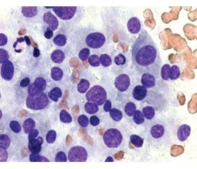
Analysis of cytological examinations of FNA smears of histologically confirmed benign focal neoplasms of the thyroid gland in Graves’ disease showed that the frequencies of cytological conclusions were distributed as follows. Cytological conclusions — nodular goiter and adenomatous node (BSRTC-2) were established in 116 cases (94.3 %). A detailed analysis of the cytological characteristics of these benign neoplasms showed that nodular and adenomatous nodes were characterized by a classic cytological picture for them, with the presence of epithelial layers of regular architecture without signs of atypia and polymorphism, with the presence of colloid or cystic degeneration. A pronounced immunocytochemical reaction of almost all epithelial cells with antibodies to Tg and TPO and the absence of CK17 was shown (Fig. 1).
Cytological signs of atypia of the follicular epithelium (BSRTC-3) were identified in 2 examined cases (2.4 %). These cases were characterized by polymorphism and signs of a certain atypia of a part of the follicular epithelium, and an intense immunocytochemical reaction of most part of the epithelium with antibodies to Tg and TPO and the absence of CK17, indicating their benign nature (Fig. 2) [14, 15].
Cytological signs of microfollicular tumor structure were identified in 5 cases (3.3 %). At the same time, a large number of microfollicular structures were found in the FNA smears, the immunocytochemical reaction with antibodies to TG and TPO was also positive in most thyrocytes, and CK17 was not detected, indicating a benign nature of tumor (Fig. 3).
In 2 of these cases, a histological diagnosis of adenoma was made, which did not contradict the cytological conclusion. In the other 3 cases, a multinodular goiter with signs of epithelial oxyphilia was histologically diagnosed.
The data of a histological examination of 409 patients with benign thyroid neoplasms who underwent thyroidectomy were analyzed. The distribution of patients with morphologically confirmed benign neoplasms by age is shown in Table 4. 
As can be seen from Table 4, the total number of cases of adenomatous goiter was 104, which accounted for 25.43 % of the total number of patients. The number of adenomas was 70 cases, of which 6 were B-cell adenomas, which accounted for 8.57 % of the total number of adenomas. Nodular hyperplasia was recorded in 141 cases 34.47 %, of which 20 cases were thyroid cysts. Solid nodules were found in 9 cases, which accounted for 2.20 % of the total number of patients, and thyroiditis — in 7 cases, which accounted for 1.71 %. Thus, among the morphologically confirmed benign neoplasms in GD, cases of nodular hyperplasia and adenomatous goiter prevailed.

Discussion 

Thus, the incidence of benign formations in GD in our study was 22.06 % of the total number — 1854 operated patients with GD. Among the group of patients, women aged 25–55 years prevailed, which accounted for 49.63 %, the second largest group was the group of patients aged 56 to 65 years — 29.58 % of the total number of patients with benign neoplasms of the thyroid gland. It should be noted that TSH-R-Ab levels of patients do not correlate with the presence of thyroid nodular neoplasms. It was found that patients with focal neoplasms were significantly (P < 0.05) older at the time of the onset of Graves’ disease than patients with diffusely altered gland without focal neoplasms.
According to cytological studies of punctates from 123 histologically confirmed benign focal neoplasms of the thyroid gland in Graves’ disease, BSRTC-2 was found in 94.3 %. At the same time, BSRTC-3 and BSRTC-4 were cytologically detected in 5.7 % of cases. In the epithelium of the FNA smears of these formations, signs of polymorphism and atypia were identified. According to the literature, Graves’ disease is characterized by polymorphism and pronounced atypia of the epithelium, and nuclear characteristics even mimic signs of papillary carcinoma [16, 17]. Incorrect cytological hyperdiagnosis in such cases was avoided by conducting additional immunocytochemical studies to determine markers that increase the accuracy of cytological diagnosis (TG, TPO, and CK17), which demonstrated the benign nature of neoplasms.
The results of the morphological study made it possible to establish that cysts/nodular hyperplasia develop mainly against the background of GD, which accounted for 34.47 % of the total number of focal formations. The second most frequent ones were adenomatous nodes, the frequency of which was 25.43 %. Thus, the study made it possible to establish the features of the cytological and pathomorphological characteristics of thyroid benign neoplasms.

Conclusions 

The incidence of benign thyroid neoplasms in Graves’ disease is 22.06 % of women aged 26 to 55 years. Cytological studies of thyroid neoplasm FNA smears in GD made it possible to clearly establish their benign nature (BSRTC-2) in 94.3 % of cases. In 5.7 % of cases, cytological categories BSRTC-3 and BSRTC-4 were identified.
Positive immunocytochemical reactions to TPO and Tg, and the absence of CK17 allow avoid cytological overdiagnosis, characteristic of benign thyroid neoplasms in GD.
Among the pathohistologically confirmed benign thyroid neoplasms in GD, in 63.82 % of cases a histological diagnosis of adenomatous or colloid goiter was established, in 34.47 % — signs of nodular hyperplasia, and in 1.71 % — the presence of thyroiditis.
 
Received 20.05.2022
Revised 06.06.2022
Accepted 08.06.2022

Список литературы

  1. Ehlers M., Schott M., Allelein S. Graves’ disease in clinical perspective. Front Biosci (Landmark Ed). 2019. 24(1). 35-47. doi: 10.2741/4708. 
  2. Wiersinga W.M. Clinical Relevance of Environmental Factors in the Pathogenesis of Autoimmune Thyroid Disease. Endocrinol. Metab. (Seoul). 2016. 31(2). 213-22. doi: 10.3803/EnM.2016.31.2.213. 
  3. Antonelli A., Ferrari S.M., Ragusa F., Elia G., Paparo S.R., Ruffilli I., Patrizio A. et al. Graves’ disease: Epidemiology, genetic and environmental risk factors and viruses. Best Pract. Res. Clin. Endocrinol. Metab. 2020. 34(1). 101387. doi: 10.1016/j.beem.2020.101387. 
  4. Rapoport B., Aliesky H.A., Chen C.R., McLachlan SM. Evidence that TSH Receptor A-Subunit Multimers, Not Monomers, Drive Antibody Affinity Maturation in Graves’ Disease. J. Clin. Endocrinol. Metab. 2015;100(6):E871-5. doi: 10.1210/jc.2015-1528. 
  5. Bartalena L., Burch H.B., Burman K.D., Kahaly G.J. A 2013 European survey of clinical practice patterns in the management of Graves’ disease. Clin. Endocrinol. (Oxf.). 2016. 84(1). 115-20. doi: 10.1111/cen.12688. 
  6. Lima P.C., Moura Neto A., Tambascia M.A., Zantut Wittmann D.E. Risk factors associated with benign and malignant thyroid nodules in autoimmune thyroid diseases. ISRN Endocrinol. 2013. 2013. 673146. doi: 10.1155/2013/673146. 
  7. Staniforth J.U.L., Erdirimanne S., Eslick G.D. Thyroid carcinoma in Graves’ disease: A meta-analysis. Int. J. Surg. 2016. 27. 118-125. doi: 10.1016/j.ijsu.2015.11.027. 
  8. Casella C., Morandi R., Verrengia A., Galani A., Molfino S., Cuka D., Groppo G., Cappelli C., Portolani N. Thyroid Cancer and Nodules in Graves’ Disease: A Single Center Experience. Endocr. Metab. Immune Disord. Drug Targets. 2021. 21(11). 2028-2034. doi: 10.2174/1871530321666201230111911. 
  9. Papanastasiou A., Sapalidis K., Goulis D.G., Michalopoulos N., Mareti E., Mantalovas S., Kesisoglou I. Thyroid nodules as a risk factor for thyroid cancer in patients with Graves’ disease: A systematic review and meta-analysis of observational studies in surgically treated patients. Clin. Endocrinol. (Oxf.). 2019. 91(4). 571-577. doi: 10.1111/cen.14069. 
  10. Khan S.H., Rather T.A., Makhdoomi R., Malik D. Nodular Graves’ disease with medullary thyroid cancer. Indian J. Nucl. Med. 2015. 30(4). 341-4. doi: 10.4103/0972-3919.164022. 
  11. Kahaly G.J. Management of Graves Thyroidal and Extrathyroidal Disease: An Update. J. Clin. Endocrinol. Metab. 2020. 105(12). 3704-20. doi: 10.1210/clinem/dgaa646. 
  12. Болгов М.Ю. Автоматизация медицинских систем. Киев: Издательство Куприянова, 2006. 464 с. 
  13. Pathology and Genetics of Tumours of Endocrine Organs. WHO Classification of Tumours. 3rd Edition. 2004. Vol. 8. Edited by DeLellis R.A., Lloyd R.V., Heitz P.U., Eng C. IARC Press, Lyon, France.
  14. Зелінська Г.В. Цитохімічне та імуноцитохімічне дослідження експресії тиреоїдної пероксидази в передопераційній діагностиці папілярного раку щитоподібної залози та його радіойодрезистентних метастазів. Онкологія. 2019. 21(3). 1-6. doi: 10.32471/oncology.2663-7928.t-21-3-2019-g.7925.
  15. Зелінська Г.В. Цитокератин 17 і тиреоїдна пероксидаза в якості імуноцитохімічних маркерів доопераційного прогнозування радіойодрезистентності та ефективності радіойодтерапії папілярного раку щитоподібної залози. Онкологія. 2019. 21(1). 31-35. doi: 10.32471/oncology.2663-7928.t-21-3-2019-g.7376. 
  16. Anderson S.R., Mandel S., LiVolsi V.A., Gupta P.K., Baloch Z.W. Can cytomorphology differentiate between benign nodules and tumors arising in Graves’ disease? Diagn. Cytopathol. 2004. 31(1). 64-7. doi: 10.1002/dc.20075. 
  17. Hang J.F., Lilo M.T., Bishop J.A., Ali S.Z. Diagnostic Accuracy of Fine Needle Aspiration in Thyroid Nodules Arising in Patients with Graves Disease. Acta Cytol. 2017. 61(2). 117-124. doi: 10.1159/000464094.

Вернуться к номеру