Інформація призначена тільки для фахівців сфери охорони здоров'я, осіб,
які мають вищу або середню спеціальну медичну освіту.

Підтвердіть, що Ви є фахівцем у сфері охорони здоров'я.


Коморбідний ендокринологічний пацієнт

Коморбідний ендокринологічний пацієнт

Международный эндокринологический журнал Том 18, №4, 2022

Вернуться к номеру

Інсулінорезистентність: метаболічні і соматичні зміни у дітей

Авторы: Gromnatska N.M., Sklyarova O.Y., Kulya O.O.
Danylo Halytsky Lviv National Medical University, Lviv, Ukraine

Рубрики: Эндокринология

Разделы: Клинические исследования

Версия для печати


Резюме

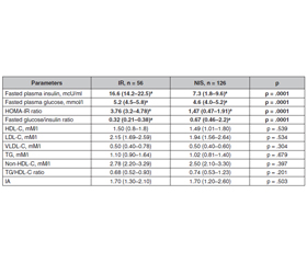
Актуальність. Інсулінорезистентність є базовим фактором етіології та патогенезу цукрового діабету 2-го типу та метаболічного синдрому і може передувати їм за багато років. Раннє виявлення початку інсулінорезистентності та пов’язаних з нею метаболічних факторів ризику у дітей запобігає розвитку цукрового діабету в дорослому житті. Мета: виявити метаболічні і соматичні зміни у дітей з інсулінорезистентністю. Матеріали та методи. З 182 дітей загальної вибірки, у яких визначено рівень базального інсуліну і глюкози, HOMA-IR та індексу глюкоза/інсулін сформовано дві групи: група 1 — 56 (30,8 %) дітей з інсулінорезистеністю, група 2 — 126 (69,2 %) дітей з нормальною чутливістю до інсуліну. Дітям проведено антропометрію, ліпідограму (загальний холестерин, тригліцериди, ХС ЛПВЩ, ХС ЛПНЩ, ХС ЛПДНЩ), лептин. Результати. З обстеженої когорти 56 дітей мали генералізоване ожиріння (ІМТ > 95-го перцентиля), 71 дитина — абдомінальне ожиріння (окружність талії > 90-го перцентиля), 55 дітей — нормальну масу тіла (ІМТ < 90-го перцентиля). Інсулінорезистентність виявлена у 21 (37,5 %) дитини з генералізованим ожирінням, 38 (39,4 %; p = 0,049) дітей з абдомінальним ожирінням і 7 (12,7 %) дітей з нормальним ІМТ (p = 0,003). У інсулінорезистентних дітей ІМТ, окружність талії і стегон були більшими, ніж в інсуліночутливих дітей. Ліпідний профіль у дітей з різною чутливістю до інсуліну не відрізнявся, проте встановлена висока кореляційна залежність ранішньої глюкози з індексом тригліцериди/ХС ЛПВЩ (r = 0,53; p = 0,001), базального інсуліну з тригліцеридами (r = 0,34; p = 0,018) та індексом тригліцериди/ХС ЛПВЩ (r = 0,54; p = 0,001). HOMA-IR корелював з ХС ЛПДНЩ (r = 0,40; p = 0,005), тригліцеридами (r = 0,49; p = 0,001), індексом тригліцериди/ХС ЛПВЩ (r = 0,43; p = 0,002). Індекс глюкоза/інсулін перебував у тісній залежності з індексом тригліцериди/ХС ЛПВЩ. Частота діагностики артеріальної гіпертензії у дітей з інсулінорезистеністю на 33,8 % перебільшувала аналогічний показник у інсуліночутливих дітей. Рівень сироваткового лептину у дітей з інсулінорезистентністю був в 1,8 раза вищим. Висновки. Інсулінорезистентність пов’язана з кардіо­метаболічними факторами ризику, такими як генералізоване та абдомінальне ожиріння, гіпертензія, дисліпідемія, гіперлептинемія і лептинорезистентність, і є біомаркером скринінгу кардіо­метаболічних захворювань.

Background. Insulin resistance is the major sign of etiology and pathogenesis of type 2 diabetes mellitus and metabolic syndrome and can precede its development for many years. Early identifying the beginning of insulin resistance in children is important to prevent diabetes mellitus in adult life. The purpose was to identify metabolic and somatic changes in children with insulin resistance. Material and methods. Out of 182 children of the general sample, who was estimated fasting plasma insulin and glucose, HOMA-IR, and glucose/insulin ratio, 2 groups were formed: group 1 — children with IR — 56 (30.8 %) and group 2 — 126 (69.2 %) children with normal insulin sensitivity. In children anthropometric data, lipid metabolism (total cholesterol, triglycerides, HDL-C, LDL-C, VLDL-C), blood pressure, leptin were determined. Results. From examined subjects 56 children were generally obese (BMI > 95th percentile), 71 children were abdominally obese (WC > 90th percentile), 55 children were with normal body mass (BMI < 90th percentile). Insulin resistance was identified in 21 (37.5 %) children with general obesity more rarely, than in 38 (39.4 %) children with abdominal obesity (p = .049) and in 7 (12.7 %) children with normal BMI (p = .003). In insulin-resistant children BMI, waist and hip circumference was larger than in children with normal insulin sensitivity. The lipid profile in children with different insulin sensitivity did not differ, but in insulin-resistant children an association of basal glucose with TG/HDL-C ratio (r = .53; p = .001), blood insulin with TG (r = .34; p = .018), and TG/HDL-C ratio (r = .54; p = .001) was estimated. The HOMA-IR significantly correlated with VLD-C (r = .40; p = .005), TG (r = .49; p = .001), TG/HDL-C ratio (r = .43; p = .002). The glucose/insulin ratio was in significant association with the TG/non-HDL-C ratio. The incidence of hypetension (> 95th percentile) diagnosis in insulin-resistant children was by 33.8 % higher (p = .001). Blood leptin concentration was 1.8 falled higher in insulin-resistant children and significantly correlates with waist circumference, fasting insulin, HOMA-IR, and diastolic blood pressure. Conclusions. Insulin resistance is related to cardiometabolic risks, such as general and abdominal obesity, hypertension, dyslipidemia, hyperleptinemia, and leptin resistance, and is a screening biomarker for children and adolescents with an increased risk of cardiometabolic diseases.


Ключевые слова

інсулінорезистентність; метаболічні фактори ризику; діти

insulin resistance; metabolic risk factors; children

Introduction

The widespread prevalence of type 2 diabetes mellitus (DM) and metabolic syndrome (MS) in the adult population and understanding that adult illnesses begin in childhood requires the early diagnosis of functional abnormalities of carbohydrate metabolism in children and their correction. Insulin resistance (IR) is one of the major signs of etiology and pathogenesis of type 2 DM and MS and can precede its development for many years [1–5]. Identifying the beginning of IR in children is important to prevent DM in the future [6].
The novel data indicate that IR and DM are common among children and adolescents [7] and increase in the children population [8]. In the meta-analysis, it was found that children’s prevalence rates of IR are between 3.1 in Greece [9] and 44 % in Pacific Island teenagers (New Zeeland) [10], with the gender higher difference in obese boys — 68.4 % [10]. However, it was also determined that a higher prevalence of IR was in girls than in boys, partly due to the difference in pubertal development [11]. 
It was estimated that changed carbohydrate metabolism and IR are estimated in 35 % children with abdominal obesity and one MS criterion, in 60.7 % children with abdominal obesity and two MS criteria and in 75 % children with abdominal obesity and three MS criteria [12].
IR was formed in the process of evolution of human beings as a way to adapt to periods of prolonged food deficit and is a physiological norm in certain periods of life such as puberty. It was demonstrated that IR occurs both as a result of primary irreversible violations and as a result of secondary reversible factors. IR etiology can be divided into acquired, hereditary, and mixed. The great majority of people with IR fall into the acquired categories [13].
IR occurs imperceptibly gradually at an early age. In the beginning, IR is asymptomatic, and metabolic and functional changes are identified only during laboratory and instrumental examinations. Type 2 DM and MS develop in the late stages of IR when it is sometimes difficult to get back the disease [13].
Insulin is an anabolic hormone that plays an important role in the regulation of glucose, lipid homeostasis, and energy storage through its natural metabolic effects on classic insulin-responsive tissues: adipose tissue, liver, and skeletal muscle. Insulin increases glucose transport across the cell membrane, by the rise of hexokinase and 6-phosphofructokinase activity, enlarges the rate of glycolysis, stimulates glycogen synthesis, increases the storage of glucose as glycogen in the liver and skeletal muscles and decreases glycogen breakdown [14, 15].
The influence of insulin on lipid metabolism is in decreasing the rate of lipolysis in adipose tissue and lowering the plasma fatty acid level, stimulation of fatty acid and triacylglycerol synthesis, increasing the uptake of triglycerides (TG) from the blood into adipose tissue and muscles, decreasing the rate of fatty acids oxidation in muscle and liver [14]. 
With IR insulin-mediated metabolic consequences on lipid and carbohydrate metabolism are changed [15]. The metabolic effect of IR can result in hyperglycemia, hypertension, dyslipidemia, visceral adiposity, elevated inflammatory markers, endothelial dysfunction [13].
Overweight and adiposity, as the result of overnutrition, are the major causative factors that induce the state of low-grade inflammation due to which accumulation of elevated levels of glucose and/or lipids in blood stream occur that leads to the activation of various transcriptional mediated molecular and metabolic pathways and IR appearance [1]. The factors determining whether or not IR develops as a result of obesity and high body mass index (BMI) are still not completely understood. 
The combination of multiple contributing factors (such as puberty, ethnic background, stress etc.) in the obese child along with incapacity of the subcutaneous fat to store excess lipid leading to intrahepatic, intramuscular, and visceral lipid deposition reduce insulin sensitivity and the development of IR and cardiovascular risk factors.
In patients who lost weight and reduce the amount of adipose tissue insulin sensitivity (IS) may return and the association of obese phenotype and type 2 DM incidence disappeare even after adjusting for HOMA-IR [16, 17]. Metabolic stress in obesity causes organelle dysfunction, especially the endoplasmic reticulum and mitochondria which regulate the glucose, fat, protein, and cholesterol metabolism. In patients with obesity occur disorders of carbohydrate and fat metabolism [18, 19].
The data of frequency of IR diagnosis, the state of carbohydrate and lipid metabolism discharge, and the nature of blood pressure, are debatable and not fully resolved which requires further study. To summarize peculiarities of metabolism and cardiovascular risk in children with IR are actual and known insufficiently.
The purpose of the study was to identify metabolic and somatic changes in children with IR.

Materials and methods

The research study was conducted at the Danylo Halytskiy Lviv National Medical University. Inclusion criteria: IR in children. Exclusion criteria: subjects suffering from infections, endocrine, and genetic disorders, consuming hormones, and suffering from secondary obesity due to other diseases.
Out of 182 children of the general sample, aged 9–18 years were involved in this research. Fasting plasma insulin and glucose, HOMA-IR, and glucose/insulin ratio was estimates and 2 groups were formed: group 1 — children and adolescents with IR — 56 (30.8 %) and group 2 — 126 (69.2 %) children and adolescents with normal insulin sensitivity (NIS). Children and adolescents of the group 1 and group 2 did not differ in age (p = .527) and gender (p = .743). The children’s age of group 1 was 15 (12–16) years, of group 2 — was 15 (12–17) years.
Before the clinical assessment, the agreement in participation in the study according to the protocol which was approved by Danylo Halytsky Lviv National Medical University Ethics Commission from parents and children was taken. A detailed medical history with a clinical examination including body mass, body mass index (BMI), neck, waist, and hip circumferences, body surface, and blood pressure (BP) were recorded.
Anthropometry was done according to standard methods. Subjects were classified as generally obese (BMI > 95 %) for sex and age. Abdominal obesity was diagnosed according to more than 90th percentile of waist circumference (WC) according to age and gender.
Glucose in serum was measured by the glucose oxidative method. Insulin was estimated by enzyme immunoassay DRG Insulin ELISA (Germany). IR was diagnosed according to HOMA-IR. It was determined that HOMA-IR is independently associated with IR, has diagnostic accuracy, and predicts IS [19]. The cut-off values used for the definition of hyperglicemia was 5.6 mM/l, IR for HOMA-IR 2.8, glucose/insulin ratio 0.48 and hyperinsulinemia 11.5 mcU/ml. 
Blood pressure (BP) was measured by mechanical manometer three times, the average data were calculated. HDL-C and TG were estimated by the enzymatic method with Cobas Integra HDL-C Test and Cobas Integra TRIGL Test (Roche Diagnostics, Switzerland), respectively.
Data statistical analysis was done by integrative systems for statistical analysis and processing Statistica 10.0 (StatSoft Inc, USA). The overall prevalence of MS and its components were estimated from the data and were associated with 95 % confidence interval. The normality of distribution was estimated according to the Shapiro-Wil-Test criterion. The results were presented as median with quartile distribution (25 and 75 percentile) and percent of the data in a group. The comparison of groups was done by using Student’s T-test and Mann-Whitney U-test depending on parametric distribution of variables. Chi-square test was used for qualitative data presented as positive/negative. For estimation of the link between qualitative characteristics correct Fisher criterion was used. The difference was significant at p-value < .05. 

Results

From 182 children of the study 56 children were generally obese (BMI > 95th percentile), 71 children were abdominally obese (WC > 90th percentile), 55 children were with normal body mass (BMI < 90th percentile). IR was identified in 21 (37.5 %) children with general obesity more rarely (p = .049), than in 38 (39.4 %) children with abdominal obesity (p = .049) and in 7 (12.7 %) children with normal BMI (p = .003) (Table 1). 
BMI in children and adolescents with IR [26.8 (21.4–28.7) kg/m2 ; p = .011] was larger than BMI in children and adolescents with NIS (24.5 (19.4–26.3 kg/m2) (Table 2). 
WC in children with IR by 9.2 % was higher than WC in children with NIS (p = .003). Hip circumference in children with IR by 8.0 % was larger than in children with NIS [93.5 (85.0–106.0) cm] (p = .048). Consequently, children and adolescents with IR were more likely to have higher BMI, WC, hip circumference than children and adolescents with NIS. No significant difference in body weight, height, neck circumference, WC /hip circumference ratio and WC/height ratio, body surface area in children with IR and NIS was observed.
It is natural that the parameters of carbohydrate metabolism in children with IR were significantly different from those in children with NIS (Table 3). Fasted plasma insulin level in children with IR was 2.3-fold, and fasted plasma glucose level was by 13.0 % higher than those in children with NIS. 
A significant difference in the HOMA-IR ratio of 2.6-fold and the fasted glucose/insulin ratio of 2.1-fold were observed in IR children compared with NIS children. The study of the correlation dependence of anthropometric parameters and carbohydrate metabolism parameters in children with IR revealed a significant association of BMI with fasted plasma glucose (r = .39; p = .023).
The lipid profile of IR children and NIR children did not differ. Though, in IR children an association of basal glucose with TG/HDL-C ratio (r = .53; p = .001), basal blood insulin with TG (r = .34; p = .018), and TG/HDL-C ratio (r = .54; p = .001) was estimated. The HOMA-IR significantly correlated with VLD-C (r = .40; p = .005), TG (r = .49; p = .001), TG/HDL-C ratio (r = .43; p = .002). The glucose/insulin ratio was in significant association with the TG/non-HDL-C ratio. 
Analysis of BP in children with different IS did not reveal the difference between systolic BP (SBP) and diastolic BP (DBP) (Table 4). 
However, the frequency of diagnosis of hypertension in IR children was by 33.8 % higher than in NIS children (p = .001). 
It was found that WC in IR children significantly correlated with the SBP (r = .29; p = .042). A highly probable correlation was observed between carbohydrate metabolism and BP: fasting insulin and HOMA-IR significantly correlated with SBP (r = .47; p = 0.001 and r = .53; p = 0.001; respectively) and DBP (r = .43; p = .002 and r =.46; p = .001; respectively). A significantly inverse correlation of the glucose/insulin ratio with SBP was observed (r = – .37; p = .008).
In IR children blood leptin concentration was 1.8 falled higher than the content of leptin in the blood of NIS children (Table 5).
In IR children essential correlation of blood leptin with CW (r = .41; p = .005), fasting insulin (r = .39; p = .007), HOMA-IR (r = .32) ; p = .031), DBP (r = .30; p = .046) was found. 

Discussion

Children with IR and NIS did not differ in age and gender, though it was estimated that children with abnormal glucose metabolism were older [20].
The range of IR was 12.7 % in children with normal body mass, 30.7 % in generally obese children, 39.4 % in abdominally obese children. Therefore, with the increase in BMI and the appearance of abdominal obesity the growth of the incidence of carbohydrate metabolism discharge was revealed [20]. It was demonstrated that regardless of the amount of total fat, the accumulation only of abdominal fat and adipose tissue dysfunction lead to an increase in adipokines and the IR appearance [21].
The study of the correlation dependence of anthropometric parameters and carbohydrate metabolism parameters in children with IR revealed a highly probable association of BMI with fasted plasma glucose (r = .39; p = .023). IR was observed to be higher with BMI increasing, which is similar to A. Ahila et al. [22]. The main visual sign in children that there is IR is the presence of general and especially abdominal obesity, in which excess adipose tissue is deposited mainly in the abdomen region. Though, the results demonstrated a notably elevated prevalence rate of IR in overweight and generally obese children [17, 23]. The same results were obtained by T. Takahashi et al. [24] that obese/overweight patients had significantly more glucose metabolism disorders than non-obese/non-overweight once. IR is usually one of the first metabolic damage diagnosed in obese children and the key risk factor for the development of comorbidities [25]. It was demonstrated that in patients with weight loss and reduction of the amount of adipose tissue NIS may return even after adjusting for HOMA-IR [16, 17].
Normally adipose tissue has a high sensitivity to insulin. In the NIS state, insulin stimulates lipid accumulation through fatty acid uptake, reesterification and lipogenesis and inhibits TG lipolysis [26]. In study of IR children an association of fasting blood glucose and insulin and HOMA-IR with the TG and TG/HDL-C ratio was revealed. That is similar to N. Çin et al. [4] who demonstrated the same positive correlation of HOMA-IR with the TG-glucose index and TG/HDL-C ratio. The Bogalusa Heart Study has shown that IR is closely related to TG levels.
Even compensatory hyperinsulinemia during long course can have a harmful influence on many tissues and, through different mechanisms, contribute to the development of other components of dyslipidemia, such as hypertriglyceridemia [27]. It was established that IR is associated with an increased lipid content and dyslipidemia [28, 29]. The obtained data prove that TG, VLDL-C, TG/HDL-C ratio can be used as IR biomarkers. 
However, the incidence of hypertension (> 95th percentile) diagnosis in IR children was 33.8 % higher than in NIS children 2 (p = .001). Insulin plays an important role in the maintenance of vascular homeostasis. Simultaneously insulin stimulates endothelial production of nitric oxide (NO), a crucial vasodilator exerting an antiaggregatory effect that prevents platelet aggregation and leukocyte adhesion to endothelial cells [30] and limiting vascular smooth muscle cells growth and migration, but mediates the release of endothelin 1, known to act as a strong vasoconstrictor. If IR appears, the balance is shifted towards mitogen-activated protein kinase/extracellular signal-regulated kinase (MAPK/ERK), which mediates inflammation, vasoconstriction, and vascular smooth muscle cell proliferation [31]. Therefore, IR coexists with endothelial dysfunction and hypertension [32].
The pathogenesis of hypertension associated even with compensatory hyperinsulinemia is stimulation in vascular smooth muscle and endothelial cells increased production of endothelin, PAI-1, proinflammatory cytokines, and an augmented surface expression of adhesion molecules [27]. IR and hyperinsulinemia cause a number of hemodynamic changes that contribute to increased BP and the development of hypertension. An association was found between fasting insulin levels and subsequent increases in BP. The main role in the development and progression of IR and related metabolic disorders is played by adipose tissue of the abdominal area, neurohormonal disorders that accompany abdominal obesity, and increased activity of the sympathetic nervous system.
In the IR children’s blood leptin concentration was 1.8 fallen higher than the content of leptin in the blood of NIS children, so with increasing blood insulin levels and the development of IR observed a proportional increase in leptinemia and the development of leptin resistance [33]. It was found that leptin mediates insulin secretion and sensitivity in peripheral tissues. Hyperleptinemia reflecting leptin resistance plays an important role in the development of IR making leptin its possible biomarker [34]. 
High correlation of leptin with basal insulin and HOMA-IR are in agreement with the findings of the studies done by P. Das et al. [35]. It was demonstrated that there is a simultaneous occurrence of IR and leptin resistance in obesity. The correlations of leptin levels with insulin, and HOMA-IR levels surely indicate the functional link between these two hormones. Leptin has an inhibitory effect on the insulin secretion, which is due to the leptin-induced proinflammatory cytokines such as C-reactive protein and Interleukin-6, causing apoptosis of pancreatic β-cells [34, 36].
It was demonstrated that leptin acts as a signal from adipocytes to pancreatic cells and reports IS. Leptin can be involved in the induction of IR by cross-reacting with insulin receptor substrates. Also, macrophage infiltration of adipose tissue and high levels of leptin in obesity lead to increased production of IL and TNF, which prevent the effects of insulin on IS tissues, and increase IR. Imbalance and dysregulation of adipokine production are involved in metabolic disorders of obesity [37]. 
In IR children was found an essential correlation of blood leptin with WC, which was in agreement with the findings of the studies done by J. Mohiti et al. stated that it was obesity leading to increased insulin and leptin levels, resulting in IR and leptin resistance [38]. Leptin levels in general increase proportionately with body fat mass.
Leptin may be the marker of obesity and IR at the same time [34], and conversely obesity is the major pathogenic factor common to both leptin resistance and IR. 

Conclusions

IR is related to cardiometabolic risks, such as general and abdominal obesity, hypertension, dyslipidemia, hyperleptinemia, and leptin resistance. 
IR is a screening marker for children and adolescents with an increased risk of cardiometabolic diseases. 
Consolidation of the actions of pediatricians and endocrinologists, new methods of early diagnostics, and improvement of the system of IR prevention are necessary. 
 
Received 19.05.2022
Revised 14.06.2022
Accepted 16.06.2022

Список литературы

  1. Rehman K., Akash M.S.H. Mechanisms of inflammatory responses and development of insulin resistance: how are they interlinked? J. Biomed. Science. 2016. 23. 87. DOI. 10.1186/s12929-0303-y.
  2. Martín-Timón I., Sevillano-Collantes C., Segura-Galindo A., Del Cañizo-Gómez F.J. Type 2 diabetes and cardiovascular disease: Have all risk factors the same strength? World J. Diabetes. 2014. 5(4). 444-70. doi: 10.4239/wjd.v5.i4.444. 
  3. Gayoso-Diz P., Otero-González A., Rodriguez-Alvarez M.X., et al. Insulin resistance (HOMA-IR) cut-off values and the metabolic syndrome in a general adult population: Effect of gender and age: EPIRCE cross-sectional study. BMC Endocr. Disord. 2013. 13. 47. DOI. 10.1186/1472-6823-13-47.
  4. Çin N.N.A., Yardımcı H., Koç N., et al. Triglycerides/high-density lipoprotein cholesterol is a predictor similar to the triglyceride-glucose index for the diagnosis of metabolic syndrome using International Diabetes Federation criteria of insulin resistance in obese adolescents: a cross-sectional study. J. Pediat. Endocrin. Metab. 2020. Published online. DOI: https://doi.org/10.1515/jpem-2019-0310.
  5. Sorokman T., Makarova O. Somatic and mental health of obese children. International Journal of Endocrinology (Ukraine). 2020. 16(7). 543-550. https://doi.org/10.22141/2224-0721.16.7.2020.219008.
  6. Bolshova O., Malinovska T. The Content of Ghrelin and Leptin in the Blood Plasma of Children and Adolescents With Hypothalamic Dysfunction. International Journal of Endocrinology (Ukraine). 2018. 14 (8). 719-24. doi:10.22141/2224-0721.14.8.2018.154849.
  7. Falalyeyeva T., Mamula Y., Scarpellini E., Leshchenko I., Humeniuk A., Pankiv I., Kobyliak N. Probiotics and obesity associated disease: an extended view beyond traditional strains. Minerva Gastroenterol. (Torino). 2021. 67(4). 348-356. doi: 10.23736/S2724-5985.21.02909-0. 
  8. Castorani V., Polidori N., Giannini C., Blasetti A., Chiarelli F. Insulin resistance and type 2 diabetes in children. Ann. Pediatr. Endocrinol. Metab. 2020. 25(4). 217-226. doi: 10.6065/apem.2040090.045. 
  9. Manios Y., Moschonis G., Kourlaba G., et al. Prevalence and independent predictors of insulin resistance in children from Crete, Greece: the Children Study. Diabet. Med. 2008. 25(1). 65-72. doi: 10.1111/j.1464-5491.2007.02318.
  10. Grant A.M., Taungapeau F.K., McAuley K.A., et al. Body mass index status is effective in identifying metabolic syndrome components and insulin resistance in Pacific Island teenagers living in New Zeeland. Metabolism: Clin. Experim. 2008. 57(4). 511-516. doi: 10.1016/j.metabol.2007.11.13.
  11. Hirscler V., Maccalini G., Karam C., et al. Age girls are insulin resistant than boys? Clin. Biochem. 2009. 42(10-11). 1051-6. doi: 10.1016/j.clinbiochem.2009.03.002.
  12. Gromnatska N., Cherkas A., Lemishko B., Kulya O. The pattern of metabolic syndrome in children with abdominal obesity. Georgian Med. News. 2019. 289. 68-72. PMID: 31215882.
  13. Petersen M.C., Shulman G.I. Mechanisms of Insulin Action and Insulin Resistance. Physiol. Rev. 2018. 98(4). 2133-2223. doi: 10.1152/physrev.00063.2017. 
  14. Czech M.P. Insulin action and resistance in obesity and type 2 diabetes. Nat. Med. 2017. 23(7). 804-814. doi: 10.1038/nm.4350. 
  15. Pankiv V.I., Yuzvenko T.Yu., Pashkovska N.V., Pankiv I.V. Effect of vitamin D on insulin resistance and anthropometric parameters in patients with type 2 diabetes mellitus. Clinical Endocrinology and Endocrine Surgery. 2019. 1(65). 53-58. DOI: http://doi.org/10.30978/CEES-2019-1-53.
  16. Soriguer F., Gutiérrez-Repiso C., Rubio-Martín E., García-Fuentes E., et al. Metabolically healthy but obese, a matter of time? Findings from the prospective Pizarra study. J. Clin. Endocrinol. Metab. 2013. 98(6). 2318-25. doi: 10.1210/jc.2012-4253. 
  17. Gepstein V., Weiss R. Obesity as the Main Risk Factor for Metabolic Syndrome in Children. Front Endocrinol. (Lausanne). 2019. 10. 568. doi: 10.3389/fendo.2019.00568. 
  18. Pankiv I.V. Intercommunication between serum vitamin D concentration and the body mass index. Obesity Reviews. 2020. 21 (Suppl. 1). EP 411. https://onlinelibrary.wiley.com/doi/epdf/10.1111/obr.13118.
  19. da Silva C.C., Zambon M.P., Vasques A.C.J., Camilo D.F., et al. Brazilian Metabolic Syndrome Study (BRAMS) Investigators. Homeostatic model assessment of adiponectin (HOMA-Adiponectin) as a surrogate measure of insulin resistance in adolescents: Comparison with the hyperglycaemic clamp and homeostatic model assessment of insulin resistance. PLoS One. 2019. 14(3). e0214081. doi: 10.1371/journal.pone.0214081.
  20. Zabarsky G., Beek C., Hagman E., Pierpont B., Caprio S., Weiss R. Impact of severe obesity on cardiovascular risk factors in youth. J. Pediatr. 2018. 192. 105-14. Doi: 10.1016/j.jpeds.2017.09.066.
  21. Hershkop K., Besor O., Santoro N., Pierpont B., Caprio S., Weiss R. Adipose insulin resistance in obese adolescents across the spectrum of glucose tolerance. J. Clin. Endocrinol. Metab. 2016. 101. 2423-31. Doi: 10.1210/jc.2016-1376.
  22. Ahila A., Vimalraj V., Raghupathy P. Insulin resistance and adverse lipid profile in obese prepubertal South Indian children. Intern. J. Pediat. Endocrinol. 2015. 78. https://doi.org/10.1186/1687-9856-2015-81-P78. 
  23. van der Aa M.P., Fazeli Farsani S., Knibbe C.A., de Boer A., van der Vorst M.M. Population-Based Studies on the Epidemiology of Insulin Resistance in Children. J. Diabetes Res. 2015. 2015. 362375. doi: 10.1155/2015/362375. 
  24. Takahashi T., Okamoto T., Sato Y., Hayashi A., Ueda Y., Ariga T. Glucose metabolism disorders in children with refractory nephrotic syndrome. Pediatr. Nephrol. 2020. 35(4). 649-657. doi: 10.1007/s00467-019-04360-1. 
  25. Mastrangelo A., Martos-Moreno G.Á., García A., Barrios V., Rupérez F.J., Chowen J.A., Barbas C., Argente J. Insulin resistance in prepubertal obese children correlates with sex-dependent early onset metabolomic alterations. Int. J. Obes (Lond). 2016. 40(10). 1494-1502. doi: 10.1038/ijo.2016.92. 
  26. Armstrong M.J., Hazlehurst J.M., Hull D., Guo K., Borrows S., Yu J., Gough S.C., Newsome P.N., Tomlinson J.W. Abdominal subcutaneous adipose tissue insulin resistance and lipolysis in patients with non-alcoholic steatohepatitis. Diabetes Obes. Metab. 2014. 16(7). 651-60. doi: 10.1111/dom.12272. 
  27. Abbasi F., Kohli P., Reaven G.M., Knowles J.W. Hypertriglyceridemia: A simple approach to identify insulin resistance and enhanced cardio-metabolic risk in patients with prediabetes. Diabetes Res. Clin. Pract. 2016. 120. 156-61. doi: 10.1016/j.diabres.2016.07.024. 
  28. Chmyr N.V., Dutka R.J. Pathogenetic and clinical features of the metabolic syndrome associated with obesity and their prognostic value. Bukovinian Med. Bullet. 2017. 23(3). 10-15. DOI: 10.24061/2413-0737/XXI.2.82.1.2017.28. (in Ukrainian)
  29. Mitsuhashi T., Hibi K., Kosuge M., et al. Relation between hyperinsulinemia and nonculprit plaque characteristics in nondiabetic patients with acute coronary syndromes. JACC Cardiov Imaging. 2011. 4(4). 392-401. DOI: 10.1016/j.jcmg.2011.02.004.
  30. Gao F., Lucke-Wold B.P., Li X., Logsdon A.F., Xu L.C., Xu S., LaPenna K.B., et al. Reduction of Endothelial Nitric Oxide Increases the Adhesiveness of Constitutive Endothelial Membrane ICAM-1 through Src-Mediated Phosphorylation. Front Physiol. 2018. 8. 1124. doi: 10.3389/fphys.2017.01124. 
  31. Boucher J., Kleinridders A., Kahn C.R. Insulin receptor signaling in normal and insulin-resistant states. Cold Spring Harb. Perspect. Biol. 2014. 6(1). a009191. doi: 10.1101/cshperspect.a009191. 
  32. Janus A., Szahidewicz-Krupska E., Mazur G., Doroszko A. Insulin Resistance and Endothelial Dysfunction Constitute a Common Therapeutic Target in Cardiometabolic Disorders. Mediators Inflamm. 2016. 2016. 3634948. doi: 10.1155/2016/3634948.
  33. Ibarra-Reynoso L.R., Pisarchyk L., Pérez-Luque E.L., Garay-Sevilla M.E., Malacara J.M. Whole-Body and Hepatic Insulin Resistance in Obese Children. PLoS ONE. 2014. 9(11). :e113576. https://doi.org/10.1371/journal.pone.0113576.
  34. Moonishaa T.M., Nanda S.K., Shamraj M., Sivaa R., Sivakumar P., Ravichandran K. Evaluation of Leptin as a Marker of Insulin Resistance in Type 2 Diabetes Mellitus. Int. J. Appl Basic Med. Res. 2017. 7(3). 176-180. doi: 10.4103/ijabmr.IJABMR_278_16. 
  35. Das P., Bhattacharjee D., Bandyopadhyay S.K., Bhattacharya G., Singh R. Association of obesity and leptin with insulin resistance in type 2 diabetes mellitus in Indian population. Indian. J. Physiol. Pharmacol. 2013. 57(1). 45-50. PMID: 24020098.
  36. Tibaldi J. Preserving insulin secretion in Type 2 diabetes mellitus. Expert Rev. Endocrinol. Metab. 2008. 3(2). 147-159. doi: 10.1586/17446651.3.2.147.
  37. Niswender K.D., Magnuson M.A. Obesity and the beta cell: lessons from leptin. J. Clin. Invest. 2007. 117(10). 2753-6. DOI: 10.1172/JCI33528.
  38. Mohiti J., Afkhami M., Babaei A. Relation between leptin and insulin in patients with type II diabetes mellitus. Int. J. Endocrinol. Metab. 2005. 3: 121-5.

Вернуться к номеру