Інформація призначена тільки для фахівців сфери охорони здоров'я, осіб,
які мають вищу або середню спеціальну медичну освіту.

Підтвердіть, що Ви є фахівцем у сфері охорони здоров'я.

Международный неврологический журнал Том 18, №3, 2022

Вернуться к номеру

Пілоцитарна астроцитома: огляд літератури

Авторы: Dipak Chaulagain, Volodymyr Smolanka, Andriy Smolanka
Regional Clinical Center of Neurosurgery and Neurology, Uzhhorod National University, Uzhhorod, Ukraine

Рубрики: Неврология

Разделы: Справочник специалиста

Версия для печати


Резюме

Пілоцитарна астроцитома (ПА) — це часта назва доброякісної пухлини мозку, яка виростає з астроцитів, клітин, що підтримують неврологічну систему. Гарві Кушинг уперше продемонстрував пілоцитарну астроцитому у 1931 році на основі серії астроцитом мозочка. Гліоми, віднесені ВООЗ до I ступеня, мають сприятливий прогноз, і пілоцитарні астроцитоми належать до цієї категорії. У осіб, які брали участь у даному дослідженні, відзначалися симптоми дефіциту функції черепно-мозкових нервів, а також симптоми атаксії та ознаки підвищення внутрішньочерепного тиску. Через зв’язок між розташуванням, розміром пухлини та наявністю супутньої гідроцефалії симптоми та ознаки ПА найчастіше спостерігаються через багато місяців. Найбільш частими симптомами є головний біль, нудота, нечіткість зору, блювання, дискомфорт у спині, підвищення внутрішньочерепного тиску та диплопія. Гістопатологічно ПА має низький або помірний рівень клітинності і складається з клітин з довгими біполярними (волосоподібними) відростками та подовженими і цитологічно м’якими ядрами, а також з ділянок з пухкою, мультиполярною (подібною до протоплазматичних астроцитів) текстурою, що складається з клітин з м’якими округло-овальними ядрами та численними короткими цитоплазматичними розширеннями. Ці ділянки багаті на волокна Розенталя. Найпоширенішим методом лікування ПА є операція з видалення пухлини. Прогноз, як правило, сприятливий, якщо пухлина повністю видалена. Якщо хірургічне втручання неможливе через розташування пухлини, дітям старшого віку та дорослим може бути корисно променеве лікування, щоб допомогти усунути будь-які залишки пухлинних клітин. Іноді використовуються хіміотерапія або інші форми таргетної терапії.

Pilocytic astrocytoma is a frequent name for a benign brain tumor that grows from astrocytes, the cells that support the neurological system. Harvey Cushing showed pilocytic astrocytoma (PA) for the first time in 1931, based on a series of cerebellar astrocytomas. Gliomas classified as grade I by the WHO have a favourable prognosis, and pilocytic astrocytomas fall within this category. There were cranial nerve deficits as well as ataxia symptoms and evidence of elevated intracranial pressure in the individuals in this investigation. Because of the relationship between the tumor’s location, size, and existence of concomitant hydrocephalus, symptoms and signs of PAs are most often seen after many months. The most frequent symptoms are headache, nausea, blurred vision, vomiting, back discomfort, elevated intracranial pressure, and diplopia. Histopathologically PA has a low to moderate level of cellularity and is composed of cells with long bipolar (hair-like) processes and elongated and cytologically bland nuclei, as well as areas with loose, multipolar (protoplasmic astrocyte-like) texture composed of cells with bland, round-to-oval nuclei and numerous short cytoplasmic extensions. These areas are rich in Rosenthal fibers. The most common treatment for PA is surgery to remove the tumor. The prognosis is generally good if the tumor is totally removed. When surgery is not an option because of the tumor’s location, older children and adults may benefit from radiation treatment to help eliminate any leftover tumor cells. Sometimes, chemo or other forms of targeted treatment are used.


Ключевые слова

астроцитома; пілоцитарна астроцитома; гліо­ма; гліома низького ступеня

astrocytoma; pilocytic astrocytoma; glioma; low grade glioma

1. Introduction

A benign brain tumor which arises from the supportive cells in the nervous system astrocytes are commonly known as pilocytic astrocytoma. Pilocytic astrocytoma is first demonstrated by Harvey Cushing in the year 1931 which was based on a sequence of cerebellar astrocytoma. PA was called as cystic cerebellar astrocytoma or juvenile pilocytic astrocytoma. This type of astrocytoma is usually low grade and commonly found in young patient, which are usually well confined. Pilocytic astrocytoma is considered as grade I gliomas by the WHO classification, which have an excellent prognosis (Knight and De Jesus, 2020). The astrocytes take the responsibilities of the functions like modulating neurotransmission, supplying nutrients to the neurons and the maintenance of the blood brain barrier. These tumors can occur in any region of the central nervous system, but usually it occurs in brainstem, hypothalamic region, cerebellum or optic nerve. As per the WHO 2016 classification these types of tumors comes under diffuse gliomas or other astrocytic tumors (Knight and De Jesus, 2020). These astrocytomas belong to the broader category of gliomas, which are tumors originating from glial cells. Pilocytic astrocytoma may also be called as low grade gliomas because the astrocytes are a type of glial cell.

2. Incidence/epidemiology/distribution of pilocytic astrocytoma

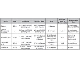
In the study of Tabash (2019) 3084 children with pilocytic astrocytoma were included whose incidence was 8227 per 1,000,000 person years. The author used Surveillance, Epidemilogy and End Results that is SEER program to obtain the children’s data who, were diagnosed with pilocytic astrocytoma between the years of 2000 and 2015. The incidence, survival and annual percentage changes (APC) were calculated. The age of the children ranged from 1 to 4 years (11.175). PA incidence has been on an upward trend for the past ten years, particularly among blacks. Furthermore it is found that infants have a poorer survival rate than adults.
Gibson, Shojaei and Susheela (n.d) made a study on astrocytoma epidemiology and demographics. Astrocytomas occur at a rate of 0.23 per 100,000 people and 700 new cases are reported every year. Approximately 148,818 people in the United States are living with brain and other nervous system cancers in 2012. According to mortality records for 2008–2012, there were 4.3 deaths per 100,000 individuals annually. Children or young adults are usually affected by the low-grade type, while adults are usually affected by the high-grade type. Approximately 62 % of all cases of pilocytic astrocytoma occur in men. Diffuse astrocytomas have a male-to-female ratio of 1.5 : 1 and anaplastic astrocytomas have a male-to-female ratio of 1.8 : 1. The caucasian race has a higher rate of astrocytoma.
A population based study was done by Burkhard et al. (2003) to determine the incidence, survival rates in the patients with pilocytic astrocytoma in canton of Zurich, Switzerland. 5.5 % of patients from 987 patients were diagnosed pilocytic astrocytoma in between the period of 1980 to 1994. The incidence rate was found to be 4.8 per 1,000,000 person years which was adjusted to the World Standard Population. The male to female ration was found to be 1.12 and the mean age was 19.6–12.7 years. 
In the study of Georgakis et al. (2016) incidence, time trends, tentative outcome disparities of childhood pilocytic astrocytoma by sociodemographic and clinical features, and survival rate of the patients diagnosed with pilocytic astrocytoma. The author used Kaplan-Meier and Cox regression analysis to examine the age standardized incidence rates. The incidence rate was 7.1 per 1 million person years. Patient’s ages ranging from less than 1 year to 14 years was involved in this study. 
Stiller et al. (2019) used population based national Registry of Childhood Tumors data to define the incidence of childhood CNS tumors. Children aging from 0 to 15 were involved in this study. The world standard population was used to calculate the age standardized rates. In 2001–10, it was 40,1 per million, age-standardised. Among the 41 %, 41 % were astrocytic tumors, 17 % were embryonal tumors, 9 % were gliomas, 7 % were epidémomas, 20 % were rare subtypes, and 5 % were unspecified tumors. In the period 1971–75 to 2006–10, the incidence of malignant and non-malignant tumors by ICD-O-3 increased by 30 and 137 %, respectively. Incidence rates in the western world were similar (table 1).

3. Symptoms/characteristics of pilocytic astrocytoma

The pilocytic astrocytoma patients may experience the following symptoms:
— headaches;
— nausea;
— vomiting;
— irritability;
— ataxia;
— vision issues.
These symptoms are caused by the increased pressure in the skull which results from the tumor. Salles et al. (2020) also witnessed the same in their study. They found that the above symptoms are due to the increase of pressure in the intracranial region. In the study of Wabbels et al. loss of unilateral visual acuity was found to be the major symptom in 70 % of adult patients and the 30 % were bilateral. Headache, neurologic deficits which includes hemiparesis and opthalmoplegia are the other symptoms encountered in their study. Collins, Jones and Giannini (2015) presented the common clinical symptoms of pilocytic astrocytoma in their study. The patients of their study experienced the symptoms of ataxia, signs of increased intracranial pressure like headache, nausea and vomiting, and also cranial nerve defects. The pilocytic astrocytoma causes loss of visual acuity or field defects when it arises in the optic pathways. Endocrine syndromes (diabetes insipidus, electrolyte imbalance or precocious puberty) may be encountered when it is developed in the hypothalamus. Hydrocephalus is caused with rapid deterioration when the tumor blocks the CSF pathways. In the study of Docampo et al. (2013) they found delayed puberty and other symptoms which are associated with the tumor’s mass effect. Chourmouzi et al. (2014), states that the symptoms and sign of pilocytic astrocytoma are commonly observed after several months of period, which are related directly to the location, size and the presence of the associated hydrocephalus. Headache, nausea, blurred vision, vomiting, neck pain, increase in intercranial pressure and diplopia are the common symptoms as per the study of Poretti, Meoded and Huisman (2012).

4. Histopathology and molecular features of PA

4.1. Histopathological features
Collins et al. (2014) states that histopathogically, PA is a low to moderately cellular tumor with compact, densely fibrillated areas rich in Rosenthal fibres, composed of cells with long bipolar (hairlike) processes and elongated cytologically bland nuclei, as well as loosely textured areas composed of multipolar cells (protoplasmic astrocyte-like), with bland, round-to-oval nuclei and multiple, relatively short cytoplasmic extensions. These regions exhibit varied degrees of mucoid background material, with microcyst formation, eosinophilic granular bodies, and hyaline droplets being prevalent. Bipolar tumor cells are frequently GFAP immunoreactive, but protoplasmic astrocyte like tumor cells are not. Areas highly similar to oligodendrogliomas may be detected in some situations, but very rarely is the oligodendroglial component prevalent. Pleomorphic nuclei, which are commonly multinucleated, can also be seen in the loose microcystic areas. Rare mitoses are acceptable, but any significant mitotic activity should raise the possibility of additional glioma diagnosis. Ki67/MIB-1 indices of up to 4 % are not uncommon. Microvascular growth is common, resulting in relatively thick-walled, hyalinized, and/or glomeruloid vessels, and infarct-like necrosis in certain cases (no pseudopalisading) (Gutman еt al., 2013). While all of these features are consistent with a PA diagnosis, they can make distinguishing PA from other gliomas challenging, especially when evaluating tiny samples. While the tumor seems rather well-defined at a macro level, variable degrees of invasion into the neighboring brain can be seen at a micro level (Ida et al., 2012). Rare cerebellar tumors form in a diffuse fashion, and molecular research may aid in recognizing these tumors as PAs.
4.2. Molecular Features
Because no prognostic indicators have been established to distinguish typical PAs from more aggressive PAs, the genetic features of the PAs have lately been examined in order to better understand and predict the behavior of this tumor (Paixao et al., 2010). Among these changes, chromosomal abnormalities (structural or numerical, as in aneuploidies), single-gene mutations, and epigenetic damage are pathways that might initiate the oncogenic molecular process.
The molecular processes of oncogenesis in PAs and diffuse astrocytomas are distinct. For example, two growth factor receptors associated with invasion and malignant development, EGFR and PDGFR, which are typically over expressed in infiltrative diffuse astrocytomas, were shown to be less expressed in PAs studied by different groups (Ji et al., 2012). Furthermore, additional genes that are changed in diffuse gliomas, including as TP53 and PTEN, are routinely expressed in PAs. Initially, no cytogenetic alterations were found in PA, and the majority of PAs had a normal karyotype, comparable to foetal astrocytes, with the bulk of the changed tumors coming from female and adult patients (Scheithauer et al., 2007). Since then, several genes have been studied. In the next sessions, we will look at the most investigated genes in PAs. 

5. Radiological features of PA

Mubarak and Naeem (2021) studied about the imaging and histpathological features of pilocytic astrocytoma which involved different regions of CNS of a series of multiple cases. In this study the authors reviewed the cases of 5 patients. As a result it is found that pilocytic astrocytomas presented a wide spectrum of neuroradiological features. Additionally to its classic appearance as a low-grade glioma, the tumor presents with an atypical presentation. Using morphological and non-morphological radiologic findings combined with site-based approaches is the most reliable method to make a preoperative diagnosis. In the study of Strong et al. (1993) the radiological images were evaluated based on their contrast enhancement degree, calcification, morphological appearance, size and the tumor distribution. Finally in the conclusion pilocytic astrocytomas can’t be predicted based on their CT and MR features, and has a poor prognosis when they behave in an aggressive manner clinically. Sharma (2022) in his study showed the radiographic features of astrocytoma. From the study it is found that a large cystic component with a brightly enhancing mural nodule, heterogeneous, mixed solid cysts, and a central necrosis, completely solid. In almost all cases, enhancement is present. A small percentage may exhibit some calcification. Hemorrhage is relatively rare.

6. Adult vs. pediatric age PA

In the study of Buckhard et al. (2003) the author states that there are many differences in the adult and pediatric pilocytic astrocytoma. Pediatric PAs most often affect the cerebellum with circumscribed, indolent, inflamed lesions. Gnekow et al. (2004) say that even progressive and recurrent pediatric PAs have been successfully treated with radiation and chemotherapy. The clinical behavior of adult PAs, however, is largely obscure because they are uncommon. The supratentorial lobar tumor occurs more frequently in adults and is typically located in the temporal or parietal region. Differences in the clinical behavior of pediatric and adult PAs have been reported inconsistently. There is some evidence that adult PAs follow a relatively benign course, similarly to pediatric PAs (Bel et al., 2004).
There is a favorable prognosis for adult patients with supratentorial PAs, both in terms of survival and neurological function, and it has been suggested that radiotherapy is not necessary following gross or subtotal resection of these lesions (Brown et al., 2004).
According to Еllis et al. (2009) adult PAs, on the other hand, are frequently found to be harmful. The recurrence rate is substantial, and tumor-related fatalities are common. Adult PAs had a recurrence incidence of 30 %, with very fast recurrence and malignant change observed in these individuals. Another research found that persistence and aggressive transformation rates in individuals who needed subsequent resection were 30 % and 50 %, respectively. As a result, investigations show that some PAs do not have a sedentary clinical course in adult patients. Adult patients exhibited the same frequency of advancement as pediatric patients in this study by Ryu et al. (2015), but those tumors that advanced did so faster and aggressively than the cases in children.
Theeler et al. (2014) studied about the clinical features and made a molecular analysis on adult pilocytic astrocytomas. Data of 127 adult patients with pilocytic astrocytoma were identified by the authors from their instituitional database. Cases with accessible tissue were examined by fluorescence in situ hybridization for BRAF-KIAA1549 fusion/duplication (B-K fusion) and submitted for mutation analysis using the Sequenom mutation profiling panel. Clinical and molecular data were used to perform subgroup analysis. Supratentorial Pas were found in most of the adult patients. 
Khan et al. (2012) made a clinical review of pediatric Pas which were treated in Pakistan at tertiary care hospital. There were 22 patients in all, with a mean age of 9.25 years. The male-to-female ratio was one to one. A symptom of elevated intracranial pressure was the most prevalent presenting characteristic. The cerebellum was the most commonly used site, followed by the cerebrum. Maximum surgical resection was performed on fifteen individuals. The male-to-female ratio was one to one. A symptom of elevated intracranial pressure was the most prevalent presenting characteristic. The cerebellum was the most commonly used site, followed by the cerebrum. Maximum surgical resection was performed on fifteen individuals. Despite the fact that there was no remaining tumor, three patients had recurrence. 

7. Pilocytic astrocytoma vs. рilomyxoid аstrocytoma

The two common types of gliomas found both in adult and children are pilocytic and pilomyxoid astrocytomas. Gliomas have a long outlook for survival since they are indolent neoplasms. These neoplasm’s genetic characteristics are well known. The pilocytic astroctytoma’s only recognized variant is the pilomyxoid asrocytoma (PMA). This was introduced in 1999 and classified as Grade II tumor by the WHO in 2007 classification. The PMA tumors commonly occur in children than in adult. When compared to pilocytic astrocytoma it shows less favorable prognosis as they exhibit cerebrospinal spread and local recurrence. As it is frequently neglected and occasionally over diagnosed among Pas, the true incidence of PMA is unknown. An initial research conducted in 1999 discovered 18 people with PAs from a large cohort of 1013 patients, yielding a rate of roughly 1.7 percent. Within a 13-year span, a single-institution research discovered that PMA accounts for roughly 10 % of patients previously identified as PA (Bhargava et al., 2013). Because of differences in diagnostic criteria, there is a considerable probability that some studies overstate the frequency of PMA within pilocytic neoplasms.
Because these tumors share identical molecular/genetic features, the PMA grading label was deleted from the 2016 WHO classification. This regrettable conclusion was based on a shaky interpretation of PMA’s biological activity. Regardless of the WHO categorization system choice, strong data probably more conclusive proof than many of the other entities in the present classification with a specified grade-shows that PMA takes a much more aggressive course than that of PA, even when age and location are accounted for (Komotar et al., 2002). Based on the existing literature data, there is sufficient evidence to support the classification of PMA as a more aggressive and higher grade variation of PA, as evidenced in well-designed and well-characterized cohort studies (Komotar et al., 2004).
Linscoft et al. (2008) made a study to expand the imaging spectrum of pilomyxoid astrocytoma. The authors stated that it is very difficult to differentiate PA and PMA based on the imaging findings alone. In comparison to PA, which is more commonly found in the posterior fossa, PMA, which is commonly found in the hypothalamus/optic chiasm, is found second most frequently intracranially. The authors saw four patients with PMAs in the cerebral hemispheres or cortical region, which is rare for PA. Intratumoral hemorrhage is the most prominent imaging feature that indicates PMA as opposed to PA. PA hemorrhage can indeed occur, but it is much less common than PMA (nearly 25 % in our series and 12 %). 
Comparison of PA and PMA of the hypothalamus/chiasm results in lower progression-free survival (26 versus 147 months) and shorter overall survival (60 versus 233 months). PMA also results in more frequent recurrence (76 % versus 50 %), often with prominent CSF dissemination. Histological confirmation of hypothalamic/chiasmatic PAs is rarely necessary. It is suggested that a neoplasm in this location, especially when it occurs with or without intratumoral hemorrhage 1 or in an older patient 2, is likely to be a PMA, not a PA. D. Chaulagain et al. (2021) suggests that more aggressive treatment may be warranted if a confirmed diagnosis of PMA is made versus PA.
Longo et al. (2016) made a study on the pilomyxoid astrocytoma of the corpus callosum presenting with primary haemorrhage in an adolescent. The authors have differentiated the diagnostic results of PMA and PA. It is found that symptoms of PMA and PA are more or less same and the rate of prognosis of PMA had high recurrence than PA. D. Chaulagain et al. (2021) made a study to express that haemorrhage is less common in PA than PMA. The authors say that Differentiating PA and PMA is a difficult task, but it is necessary because of prognostic factors. PMA can be more likely to be diagnosed when certain imaging features, including hemorrhagic component, solid mass, leptomeningeal dissemination, and greater T2-signal intensity inside the lesion, are present. 
Ho et al. (2019) made a comparison of PA and PMA using dynamic susceptibility contrast perfusion and diffusion weighted imaging. 49 patients were involved in this study where 30 patients were with infratentorial pilocystic astrocytoma, 8 with supratentorial pilocystic astrocytoma, 6 with supratentorial pilomyxoid astrocytoma, and 5 with infratentorial pilomyxoid astrocytoma. 

8. Pilocytic аstrocytoma vs. рleomorphic xanthoastrocytoma

Pleomorphic xanthoastrocytoma (PXA) is an uncommon, benign tumor in the brain that most likely develops from astrocytes, which are nervous system cells that form the brain’s supporting network. Pleomorphic xanthoastrocytoma is most commonly found in the cerebral hemisphere, the topmost portions of the brain, and the leptomeninges, one of the layers that surround the brain. It only occurs on rare occasions in the spinal cord. Males and females are equally affected by pleomorphic xanthoastrocytomas, and the median age at diagnosis is 12 years. A PXA will seldom develop into a more malignant tumor. 
According to Shaikh et al. (2019) рleomorphic xanthoastrocytoma (PXA) is an astrocytic tumor that is hypothesised to be caused by subpial astrocytes or their progenitors. It is uncommon, accounting for about 1 % of all astrocytomas. Kepes created the acronym PXA in 1979, after it was first characterised in 1973. It was formally classified as a WHO grade 2 tumor in the WHO classification system of malignancies of the central nervous system (CNS) in 1993. Anaplastic grade 3 forms are described further on. The name PXA, while representing a single histologic entity, is likely to reflect more than one tumor type as described molecularly. 

9. Surgical treatment of adult pilocytic astrocytoma

Adib et al. (2021) made a study on the surgical management of primary and secondary pilocytic astrocytoma of the cerebellopontine angle. The author identified the patients who undergone the PA-CPA surgery from January 2004 to 2019 December through the medical file of their neurosurgery department computer data. According to our criteria, three patients were placed under general anaesthesia in the supine position and one in the semisitting position. In the instance of semisitting posture, the anesthesiologic setup incorporated transesophageal echocardiography for early identification of air emboli. A retrosigmoid craniectomy was performed on all patients. Intraoperative monitoring comprised electromyography (EMG) and motor evoked potentials (MEP) observations of the facial nerve and lower cranial nerves, as well as auditory evoked potentials, MEP, and sensory generated possibilities of the upper and lower limbs. 
Kayama, Tominaga and Yashimoto (1996) made a study to clarify treatment recommendations for pilocytic astrocytoma patients. They examined the records of 41 individuals who had a histologic diagnosis of pilocytic astrocytoma at the time when CT imaging was available to detect recurrence. The treatment recommendations were based on outcomes studies relating to patient age, tumor location, surgical treatment, and radiation therapy. Radiation therapy administered following surgery inhibited any remaining tumor. We came to the conclusion that the optimal therapy for pilocytic astrocytoma is 1) complete excision, if feasible, followed by 2) irradiation of any remaining tumor to prevent recurrence.
In the study of Apanisile and Karosi (2017) surgical management of pilocytic astrocytoma of the optic nerve is reported. A preoperative MRI scan indicated a soft tissue mass surrounding the right eye’s retrobulbar region with undamaged orbital bony walls. It was separated easily from the muscles and detached from the optic nerve and the globe during surgery. Histopathologic examination showed the presence of a benign astrocytoma. The follow-up exam found no evidence of recurrence or persistent tumor. The authors conclude that unilateral optic nerve astrocytoma surgical intervention is a generally safe method that allows for total or partial tumor excision with minimum morbidity and a low recurrence rate.
When possible, surgery to remove the tumor is the usual therapy for PA. If the tumor is completely eliminated, the prognosis is typically favorable. If the tumor is in a location where it cannot be completely removed during surgery, adults and older children may be given radiation therapy to help destroy any remaining tumor cells. Chemotherapy and/or targeted treatment are sometimes employed. Chemotherapy may be indicated following surgery in younger children, although radiation is rarely utilized. This is due to the fact that radiation can create long-term growth and development issues in young children. Because PAs grow extremely slowly, your doctor may choose to wait and see if the tumor returns following surgery. This is because to the fact that the dangers of radiation therapy may outweigh the chance of the PA re-growing. This is possible for both adults and children.

Conclusions

The cerebellum is the most prevalent location for pilocytic astrocytomas, which are the most common childhood brain tumors in the general population. Even while total excision is the treatment of choice for the vast majority of malignancies, recurrence is not unheard of in certain cases.
 
Received 25.03.2022
Revised 09.04.2022
Accepted 15.04.2022

Список литературы

  1. Yonekawa Y., Lutolf U.M., Kleihues P., Ohgaki H. A population-based study of the incidence and survival rates in patients with pilocytic astrocytoma. J. Neurosurg. 2003. 98. 1170-1174.
  2. Brown P.D., Buckner J.C., O’Fallon J.R., Iturria N.L., Brown C.A., O’Neill B.P., Scheithauer B.W., Dinapoli R.P., Arusell R.M., Abrams R.A., Curran W.J., Shaw E.G., North Central Cancer Treatment Group, Mayo Clinic. Adult patients with supratentorial pilocytic astrocytomas: a prospective multicenter clinical trial. Int. J. Radiat. Oncol. Biol. Phys. 2004. 58. 1153-1160.
  3. Ellis J.A., Waziri A., Balmaceda C., Canoll P., Bruce J.N., Sisti M.B. Rapid recurrence and malignant transformation of pilocytic astrocytoma in adult patients. J. Neuro-Oncol. 2009. 95. 377-382.
  4. Bell D., Chitnavis B.P., Al-Sarraj S., Connor S., Sharr M.M., Gullan R.W. Pilocytic astrocytoma of the adult — clinical features, radiological features and management. Br. J. Neurosurg. 2004. 18. 613-616.
  5. Gnekow A.K., Kortmann R.D., Pietsch T., Emser A. Low grade chiasmatic-hypothalamic glioma-carboplatin and vincristin chemotherapy effectively defers radiotherapy within a comprehensive treatment strategy — report from the multicenter treatment study for children and adolescents with a low grade glioma — HIT-LGG 1996 — of the Society of Pediatric Oncology and Hematology (GPOH). Klin. Padiatr. 2004. 216. 331-342.
  6. Bhargava D., Sinha P., Chumas P., Al-Tamimi Y., Shivane A., Chakrabarty A., et al. Occurrence and distribution of pilomyxoid astrocytoma. Br. J. Neurosurg. 2013. 27. 413-8.
  7. Komotar R.J., Burger P.C., Goldthwaite P.T., Tihan T. Hypothalamic-Chiasmatic Astrocytomas with Pilocytic and Pilomyxoid Morphology: A Reappraisal of 63 Cases. Neurosurgery. 2002. 51. 552-3.
  8. Komotar R.J., Burger P.C., Carson B.S., Brem H., Olivi A., Goldthwaite P.T., et al. Pilocytic and pilomyxoid hypothalamic/chiasmatic astrocytomas. Neurosurgery. 2004. 54. 72-9.
  9. Adib S., Schuhmann M., Hempel J., Bornemann A., Zamora R., Tatagiba M. Surgical management of primary and secondary pilocytic astrocytoma of the cerebellopontine angle (in adults and children) and review of the literature. Neurosurgical. Review. 2020. 44(2). 1083-1091.
  10. Apanisile I., Karosi T. Surgical Management of Pilocytic Astrocytoma of the Optic Nerve: A Case Report and Review of the Literature. Case Reports in Oncological Medicine. 2017. 2017. 1-7.
  11. Burkhard C., Di Patre P., Schüler D., Schüler G., Yaşargil M., Yonekawa Y., Lütolf U., Kleihues P., Ohgaki H. A population-based study of the incidence and survival rates in patients with pilocytic astrocytoma. Journal of Neurosurgery. 2003. 98(6). 1170-1174.
  12. Chourmouzi D., Papadopoulou E., Konstantinidis M., Syrris V., Kouskouras K., Haritanti A., Karkavelas G., Drevelegas A. Manifestations of pilocytic astrocytoma: a pictorial review. Insights into Imaging. 2014. 5(3). 387-402.
  13. Georgakis M., Karalexi M., Kalogirou E., Ryzhov A., Zborovskaya A., Dimitrova N., et al. Incidence, time trends and survival patterns of childhood pilocytic astrocytomas in Southern-Eastern Europe and SEER, US. Journal of Neuro-Oncology. 2016. 131(1). 163-175.
  14. Kayama T., Tominaga T., Yoshimoto T. Management of pilocytic astrocytoma. Neurosurgical Review. 1996. 19(4). 217-220.
  15. Chaulagain D., Smolanka V.I., Smolanka A.V., Havryliv T.S. The impact of extent of resection in surgical outcome of pilomyxoid astrocytoma: a case study. Ukrainian Neurosurgical Journal. 2021. 27(4). 43-48. https://doi.org/10.25305/unj.242926.
  16. Mair M., Wöhrer A., Furtner J., Simonovska A., Kiesel B., Oberndorfer S., et al. Clinical characteristics and prognostic factors of adult patients with pilocytic astrocytoma. Journal of Neuro-Oncology. 2020. 148(1). 187-198.
  17. Mwita C., Koech F., Sisenda T., Patel K., Macharia B., Rahangdale D. Clinicopathologic Features and Early Surgical Outcome of Astrocytomas in Eldoret, Kenya. Journal of Neurosciences in Rural Practice. 2018. 09(03). 363-369.
  18. Pompili A., Caperle M., Pace A., Ramazzotti V., Raus L., Jandolo B., Occhipinti E. Quality-of-life assessment in patients who had been surgically treated for cerebellar pilocytic astrocytoma in childhood. Journal of Neurosurgery. 2002. 96(2). 229-234.
  19. Stiller C., Bayne A., Chakrabarty A., Kenny T., Chumas P. Incidence of childhood CNS tumors in Britain and variation in rates by definition of malignant behaviour: population-based study. BMC Cancer. 2019. 19(1).
  20. Tabash M. Characteristics, survival and incidence rates and trends of pilocytic astrocytoma in children in the United States; SEER-based analysis. Journal of the Neurological Sciences. 2019. 400. 148-152.
  21. Theeler B., Ellezam B., Sadighi Z., Mehta V., Tran M., Adesina A., Bruner J., Puduvalli V. Adult pilocytic astrocytomas: clinical features and molecular analysis. Neuro-Oncology. 2014. 16(6). 841-847.
  22. Voronina N., Aichmüller C., Kolb T., Korshunov A., Ryzhova M., Barnholtz-Sloan J., et al. The age of adult pilocytic astrocytoma cells. Oncogene. 2021. 40(16). 2830-2841.
  23. Becker A.P, Scapulatempo-Neto C., Neder L., Chimelli L., Reis R.M. Clinical management and evolving novel therapeutic strategies for patients with brain tumors. Intechopen. 2013. 127-143.
  24. Collins V.P., Jones D.T.W., Giannini C. Pilocytic astrocytoma: pathology, molecular mechanisms and marker. Acta Neuropathol. 2015.
  25. Salles D., Laviola G., Malinverni A.C., Stávale J.N. Pilocytic astrocytoma: A review of general, clinical, and molecular characteristics. Journal of Child Neurology. 2020. 35(12). 852-858. https://doi.org/10.1177/0883073820937225. 
  26. Koeller K.K., Rushing. E.J. Pilocytic astrocytoma: radiologic-pathologic correlation. Radiographics. 2004. 24(6). 1693-1708.
  27. Strong J.A., Hatten H.P., Brown M.T., Debatin J.F., Friedman H.S., Oakes W.J., Tien R. Pilocytic Astrocytoma: Correlation between the initial imaging features an clinical aggressiveness. AJR. 1992. 1993(161). 369-372.
  28. Mubarak F., Naeem A. Imaging and Histopathological Features of Pilocytic Astrocytoma Involving Various Locations of Central Nervous System — Series of Multiple Cases. Journal of Neurophysiology and Neurological Disorders. 2021. 9(101). 1-7.
  29. Ryu H., Jung T., Lee G., Lee K., Jung S., Jung S., Baek H. Differnces in the clinical couces of pediatric and adult pilocytic astrocytomas with progression: a single-instituition study. Childs Nerv. System. 2015.
  30. Khan M.A., Godil S.S., Tabani H., Panju S.A., Enam S.A. Clinical review of pediatric pilocytic astrocytomas treated at a tertiary care hospital in Pakistan. Surgicval Neurology International. 2012. 3(90).
  31. Ding C., Tihan T. Recent progress in the pathology and genetics of pilocytic and pilomyxoid astrocytomas. Balken. Med. J. 2019. 36(2019). 3-11.
  32. Chaulagain D., Smolanka V., Smolanka A. Diagnosis and management of astrocytoma: a literature review. International Neurological Journal. 2022. 18(1). 23-29. https://doi.org/10.22141/2224-0713.18.1.2022.925.
  33. Longo M., Adams Perez J., Oliveira F., Antunes A., Vedolin L., Duarte J.A. Pilomyxoid astrocytoma of the corpus callosum presenting with primary haemorrhage in an adolescent. BJR Case Rep. 2010. 2. 20150020.
  34. Kleinschmidt-DeMasters B.K., Donson Andrew M., Richmond Abby M., Pekmezci Melike, Tihan Tarik, Foreman Nicholas K. SOX10 Distinguishes Pilocytic and Pilomyxoid Astrocytomas From Ependymomas but Shows no Differences in Expression Level in Ependymomas From Infants Versus Older Children or Among Molecular Subgroups. Journal of Neuropathology & Experimental Neurology. 2016. nlw010. doi: 10.1093/jnen/nlw010. 
  35. Ho Chang Y., Supakul Nucharin, Patel Parth U., Seit Vetana, Groswald Michael, Cardinal Jeremy, Lin Chen, Kralik Stephen F. Differentiation of pilocytic and pilomyxoid astrocytomas using dynamic susceptibility contrast perfusion and diffusion weighted imaging. Neuroradiology. 2019. doi: 10.1007/s00234-019-02310-0.

Вернуться к номеру