Резюме
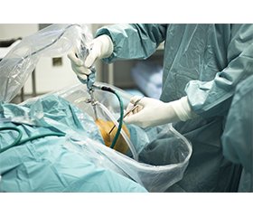
Актуальність. Остеоартроз (ОА) колінного суглоба — прогресуюче невиліковне захворювання, яке у разі тяжкого перебігу призводить до тотального ендопротезування суглоба, яке потребує значних економічних витрат та медико-соціальної адаптації і має певну кількість післяопераційних ускладнень та несприятливих результатів. Тому останнім часом особлива увага приділяється профілактиці та лікуванню ранніх стадій захворювання. Мета: провести системний аналіз наукової літератури щодо перспективи та можливостей використання артроскопії при ранній діагностиці моногонартрозу. Матеріали та методи. У базах даних PubMed та Medline за період 2010–2024 рр. було проведено літературний пошук, для якого використовували такі медичні предметні рубрики і ключові слова: деформуючий остеоартроз колінного суглоба, остеоартроз колінного суглоба, односторонній деформуючий остеоартроз колінного суглоба, односторонній остеоартроз колінного суглоба, гонартроз, моногонартроз, артроскопія, діагностика, лікування, deforming osteoarthritis of the knee joint, osteoarthritis of the knee joint, unilateral deforming osteoarthritis of the knee joint, unilateral osteoarthritis of the knee joint, gonarthrosis, monogonarthrosis, arthroscopy, diagnosis, treatment. За необхідності в окремих випадках використовували джерела літератури, що виходять за межі періоду пошуку. Загальний пошук виявив 48 джерел щодо використання артроскопії як діагностично-лікувального методу на ранніх стадіях моногонартрозу. Первинне виключення стосувалось літературних джерел, у яких артроскопія застосовувалася в діагностиці та лікуванні пізніх стадій остеоартрозу колінного суглоба (n = 38). До вторинного виключення були віднесені літературні джерела, які містили тільки довідкову інформацію (резюме, рисунки, перелік літератури) (n = 12). У результаті залишилися тільки релевантні повнотекстові статті у фахових журналах (n = 15). Результати. Відсутність кореляції між клінічною симптоматикою та рентгенологічними ознаками ОА колінного суглоба обумовлює низьку доступність ортопедичної допомоги: більше ніж 30 % хворих з уперше встановленим діагнозом мають виражену стадію захворювання, а в низці випадків патологія діагностується лише у зв’язку з проявом ускладнення; діагноз остеоартрозу через велику частку безболісного розвитку захворювання (40 %) встановлюється часто на термінальних стадіях. Усе це свідчить про необхідність подальших досліджень різних факторів, що впливають на частоту, поширеність, економічний та соціальний тягар остеоартрозу колінного суглоба. Артроскопія потенційно є золотим стандартом для перевірки неінвазивних методів оцінки, як-от магнітно-резонансна томографія, оскільки вона забезпечує сильне збільшення та прямий огляд суглобового хряща з неруйнівною інтерактивною оцінкою його структури та функціональних властивостей. Артроскопія дозволяє зробити більш докладний опис глибини та поширеності уражень, а також виявити тонкі зміни, як-от розм’якшення хряща, фібриляції та тангенціальне лущення. Клінічна симптоматика та структурні зміни елементів колінного суглоба, візуалізовані під час артроскопії у пацієнтів з моногонартрозом, висвітлюються у поодиноких дослідженнях, частину яких було опубліковано 10 років тому. Результати сучасних артроскопічних досліджень можуть стати важливим внеском в розробку діагностичних та диференційно-діагностичних критеріїв ранніх стадій моногонартрозу. Висновки. Встановлено на основі інформаційно-аналітичних досліджень сучасної наукової літератури, що остеоартроз колінного суглоба супроводжується стійким болем, суттєвим обмеженням функції нижньої кінцівки, зниженням працездатності, що нерідко призводить до ендопротезування суглоба. Діагностика ОА на ранніх стадіях утруднена внаслідок відсутності патогномонічних клінічних, рентгенологічних й лабораторних показників, а у разі моногонартрозу з синовітом ускладнюється через диференціацію зі специфічними артритами колінного суглоба. Артроскопія дозволяє виконати необхідний обсяг діагностично-лікувальних заходів з верифікацією патологічного процесу й визначенням стадії гонартрозу.
Background. Knee osteoarthritis is a progressive incurable disease that in severe cases leads to total joint replacement, which requires significant economic costs and medical and social adaptation, has a number of postoperative complications and adverse outcomes. Therefore, special attention has recently been paid to the prevention and treatment of the early stages of the disease. The purpose of the study was to conduct a systematic analysis of scientific literature on the prospects and possibilities of using arthroscopy in the early diagnosis of monoarthrosis. Material and methods. A literature search was conducted in the PubMed and MEDLINE databases for 2010–2024 using the following medical subject headings and keywords: “deforming osteoarthritis of the knee joint”, “osteoarthritis of the knee joint”, “unilateral deforming osteoarthritis of the knee joint”, “unilateral osteoarthritis of the knee joint”, “gonarthrosis”, “monoarthrosis”, “arthroscopy”, “diagnosis”, “treatment”. If necessary, literature sources beyond the search period were used in some cases. A general search revealed 48 references on the use of arthroscopy as a diagnostic and therapeutic method in the early stages of monoarthrosis. The primary exclusion concerned the literature in which arthroscopy was used for the diagnosis and treatment of late-stage knee osteoarthritis (n = 38). The secondary exclusion included literature sources that contained only background information (summary, figures, references) (n = 12). As a result, only relevant full-text articles in professional journals remained (n = 15). Results. The lack of correlation between clinical symptoms and radiological signs of knee osteoarthritis causes low availability of orthopaedic care: more than 30 % of newly diagnosed patients have a severe stage of the disease, and in some cases the pathology is detected only in connection with the manifestation of complications; the diagnosis of osteoarthritis due to a large percentage of painless development of the disease (40 %) is often established at terminal stages. All of this suggests the need for further research into the various factors that influence the frequency, prevalence, economic and social burden of knee osteoarthritis. Arthroscopy is potentially the gold standard for validating non-invasive assessment methods such as magnetic resonance imaging, as it provides high magnification and direct view of articular cartilage with non-destructive interactive assessment of its structure and functional properties. Arthroscopy allows for a more detailed description of the depth and extent of lesions, as well as the detection of subtle changes such as cartilage softening, fibrillations, and tangential peeling. Clinical symptoms and structural changes in the knee joint elements visualised during arthroscopy in patients with monoarthrosis are covered in a few studies, some of which were published 10 years ago. The results of modern arthroscopic studies can be an important contribution to the development of diagnostic and differential diagnostic criteria for the early stages of monoarthrosis. Conclusions. Based on information and analytical studies of modern scientific literature, it has been found that knee osteoarthritis is accompanied by persistent pain, significant limitation of the lower limb function, and reduced ability to work, which often leads to joint replacement. Diagnosis of osteoarthritis in the early stages is difficult due to the absence of pathognomonic clinical, radiological and laboratory parameters, and in case of monoarthrosis with synovitis, it is complicated by differentiation with specific arthritis of the knee joint. Arthroscopy allows performing the necessary scope of diagnostic and therapeutic measures with verification of the pathological process and determination of gonarthrosis stage.
Список литературы
1. Борткевич О.П., Гармаш О.О., Калашніков О.В., Коваленко В.М., Полулях М.М., Проценко Г.О. та ін. Клінічна настанова [Остеоартроз. Клінічні рекоменадації]. Київ, 2017. 481 с. [Електронний ресурс]. Режим доступу: https://www.dec.gov.ua/wp-content/uploads/2019/11/akn_osteo.pdf.
2. Інтернет-ресурс Державної служби статистики України [Інтернет-ресурс Державної статистичної служби України] [Електронний ресурс]. Режим доступу: http://database. ukrcensus.gov.ua/MULT/Dialog/statfile_c.asp.
3. Колесніченко В.А., Голка Г.Г., Ханик Т.Я., Веклич В.М. Епідеміологія остеоартрозу колінного суглоба. The Journal of VN Karazin Kharkiv National University. 2021;(43):115-126.
4. Abraham S, Patel S. Monoarticular Arthritis. [Updated 2021 Aug 27]. In: StatPearls [Internet]. Treasure Island (FL): StatPearls Publishing; 2022 Jan. Available from: https://www.ncbi.nlm.nih.gov/books/NBK542164/
5. Altuwairqi AA, Qronfla HM, Aljehani LS, et al. The Association Between Gonarthrosis Pain Severity and Radiographic Findings on X-Ray: A Cross-Sectional Study. Cureus. 2023 Feb 21;15(2):e35258. doi: 10.7759/cureus.35258.
6. Arthritis Foundation. Arthritis by the Numbers. In: Atlanta, GA: Arthritis Foundation; 2019: https://www.arthritis.org/Documents/Sections/About-Arthritis/arthritis-facts-stats-figures.pdf. Accessed April 5, 2019.
7. Astephen Wilson JL, Kobsar D. Osteoarthritis year in review 2020: mechanics. Osteoarthritis and Cartilage. 2021;29:161-169. https://doi.org/10.1016/j.joca.2020.12.009.
8. Bannuru RR, Osani MC, Vaysbrot EE, Arden NK, Bennell K, Bierma-Zeinstra SMA, et al. OARSI guidelines for the non-surgical management of knee, hip, and polyarticular osteoarthritis. Osteoarthritis Cartilage. 2019;27(11):1578-89. Doi: https://doi.org/10.1016/j.joca.2019.06.011.
9. Barr AJ, Campbell TM, Hopkinson D, Kingsbury SR, Bowes MA, Conaghan PG. A systematic review of the relationship between subchondral bone features, pain and structural pathology in peripheral joint osteoarthritis. Arthritis Res & Therapy. 2015;17:228. DOI: 10.1186/s13075-015-0735-x.
10. Becker JA, Daily JP, Pohlgeers KM. Acute Monoarthritis: Diagnosis in Adults. Am Fam Physician. 2016 Nov 15;94(10):810-816.
11. Berenbaum F, Wallace IJ, Lieberman DE, Felson DT. Modern-day environmental factors in the pathogenesis of osteoarthritis. Nat. Rev. Rheumatol. 2018;14:674-681.
12. Berteau J-P. Knee Pain from Osteoarthritis: Pathoge-nesis, Risk Factors, and Recent Evidence on Physical The-rapy Interventions. J. Clin. Med. 2022;11:3252. https:// doi.org/10.3390/jcm11123252.
13. Bliddal H, Leeds AR, Christensen R. Osteoarthritis, obesity and weight loss: evidence, hypotheses and horizons — a scoping review. Obes Rev. 2014 Jul;15(7):578-586. Doi: https://doi.org/10.1111/obr.12173.
14. Bruyere O, Honvo G, Veronese N, Arden NK, Branco J, Curtis EM, Al-Daghri NM [et al.]. An updated algorithm recommendation for the management of knee osteoarthritis from the European Society for Clinical and Economic Aspects of Osteoporosis, Osteoarthritis and Musculoskeletal Diseases (ESCEO). Semin Arthritis Rheum. 2019 Dec;49(3):337-350. doi: 10.1016/j.semarthrit.2019.04.008.
15. Chamorro-Moriana G, Perez-Cabezas V, Espuny-Ruiz F, Torres-Enamorado D, Ridao-Fernandez C. Assessing knee functionality: Systematic review of validated outcome measures. Annals Phys Rehab Med. 2022;65:101608. https://doi.org/10.1016/j.rehab.2021.101608.
16. Chen D, Shen J, Zhao W, Wang T, Han L, Hamilton JL, Im H-J. Osteoarthritis: toward a comprehensive understanding of pathological mechanism. Bone Research. 2017;5:16044. Doi: https://doi.org/10.1038/boneres.2016.44.
17. Chilelli BJ, Cole BJ, Farr J, Lattermann C, Gomoll AH. The Four Most Common Types of Knee Cartilage Damage Encountered in Practice: How and Why Orthopaedic Surgeons Manage Them. In: AAOS Instr Course Lect. 2017;66:507-530.
18. Cooper C, Bruyere O, Arden N, Branco J, Brandi ML, Herrero-Beaumont G, Berenbaum F [et al.]. Can we identify patients with high risk of osteoarthritis progression who will respond to treatment? A focus on epidemiology and phenotype of osteoarthritis. Drugs Aging. 2015 Mar;32(3):179-87. doi: 10.1007/s40266-015-0243-3.
19. Cui A, Li H, Wang D, Zhong J, Chen Y, Lu H. Glo-bal, regional prevalence, incidence and risk factors of knee osteoarthritis in population-based studies. EClinicalMedicine. 2020;2930:100587. Doi: https://doi.org/10.1016/j.eclinm.2020.100587.
20. Demehri S, Guermazi A, Kwoh CK. Diagnosis and longitudinal assessment of osteoarthritis: review of available imaging techniques. Rheum Dis Clin North Am. 2016;42:607-20.
21. de Oliveira Vargas e Silva NC, dos Anjos RL, Santana MMC, Battistella LR, Alfieri1 FM. Discordance between radiographic findings, pain, and superficial temperature in knee osteoarthritis. Reumatologia. 2020;58,6:375-380. DOI: https://doi.org/10.5114/reum.2020.102002.
22. Emery CA, Whittaker JL, Mahmoudian A, Lohmander LS, Roos EM, Bennell KL, et al. Establishing outcome measures in early knee osteoarthritis. Nat. Rev. Rheumatol. 2019;15:438-448.
23. Finney A, Dziedzic KS, Lewis M, Healey E. Multisite peripheral joint pain: a cross-sectional study of prevalence and impact on general health, quality of life, pain intensity and consultation behaviour. BMC Musculoskeletal Disorders. 2017;18:535. Doi: https://doi.org/10.1186/s12891-017-1896-3.
24. Gorial FI, Anwer Sabah SA, Kadhim MB, Jamal NB. Functional Status in Knee Osteoarthritis and its Relation to Demographic and Clinical Features. Mediterr J Rheumatol. 2018;29(4):207-10.
25. Guo J, Huang X, Dou L, et al. Aging and aging-related diseases: from molecular mechanisms to interventions and treatments. Sig Transduct Target Ther. 2022;7:391. https://doi.org/10.1038/s41392-022-01251-0.
26. Harkey MS, Davis JE, Lu B, Price LL, Ward RJ, Mac-kay JW, et al. Early pre-radiographic structural pathology precedes the onset of accelerated knee osteoarthritis. BMC Musculoskelet. Disord. 2019;20:1-10.
27. Huang Y, Deng W, Zheng S, et al. Relationship between monocytes to lymphocytes ratio and axial spondyloarthritis. Int Immunopharmacol. 2018;57:43-46. doi: 10.1016/j.intimp.2018.02.008.
28. Hunter DJ, Zhang W, Conaghan PG, Hirko K, Menashe L, Li L, Reichmann WM, Losina E. Systematic review of the concurrent and predictive validity of MRI biomarkers in OA. Osteoarthritis Cartilage. 2011 May;19(5):557-588. doi: 10.1016/j.joca.2010.10.029.
29. Jones KJ, Sheppard WL, Arshi A, Hinckel BB, Sherman SL. Articular Cartilage Lesion Characteristic Reporting Is Highly Variable in Clinical Outcomes Studies of the Knee. Cartilage. 2019 Jul;10(3):299-304. doi: 10.1177/1947603518756464.
30. Kelli D Allen, Yvonne M Golightl. Epidemiology of osteoarthritis: state of the evidence. Curr Opin Rheumatol. 2016, 27:000-000. Doi: https://doi.org/10.1097/BOR.0000000000000161.
31. Kim S-H, Park K-N. Evaluation of the Relationships Between Kellgren-Lawrence Radiographic Score and Knee Osteoarthritis-related Pain, Function, and Muscle Strength. Phys. Ther. Korea 2019;26(2):69-75. https://doi.org/10.12674/ptk.2019.26.2.069.
32. Kloppenburg M, Berenbaum F. Osteoarthritis year in review 2019: Epidemiology and therapy. Osteoarthr. Cartil. 2020;28:242-248.
33. Krakowski P, Karpinski R, Jojczuk M, Nogalska A, Jonak J. Knee MRI Underestimates the Grade of Cartilage Lesions. Appl. Sci. 2021;11:1552. https://doi.org/10.3390/ app11041552.
34. Krishnasamy P, Hall M, Robbins SR. The role of skeletal muscle in the pathophysiology and management of knee osteoarthritis. Rheumatology. 2018 May;57,4:iv22-iv33. https://doi.org/10.1093/rheumatology/kex515.
35. Li D, Li S, Chen Q, Xie X. The Prevalence of Symptomatic Knee Osteoarthritis in Relation to Age, Sex, Area, Region, and Body Mass Index in China: A Systematic Review and Meta-Analysis. Front. Med. 2020 July16. Doi: https://doi.org/10.3389/fmed.2020.00304.
36. Liu C, Wan Q, Zhou W, Feng X, Shang S. Factors associated with balance function in patients with knee osteoarthritis: An integrative review. Int J Nursing Sciences. 2017;4(4):402-409. https://doi.org/10.1016/j.ijnss.2017.09.002.
37. Mathiessen A, Conaghan PG. Synovitis in osteoarthritis: current understanding with therapeutic implications. Arthritis Res Ther. 2017;19(1):18-21. https://doi.org/10.1186/s13075-017-1229-9.
38. Mohajer B, Dolatshahi M, Moradi K, Najafzadeh N, Eng J, Zikria B, et al. Role of Thigh Muscle Changes in Knee Osteoarthritis Outcomes: Osteoarthritis Initiative Data. Radiology. 2022;305(1):169-178. https://doi.org/10.1148/radiol.212771.
39. Mora JC, Przkora R, Cruz-Almeida Y. Knee osteoarthritis: pathophysiology and current treatment modalities. Journal of Pain Research. 2018;11:2189-2196. http://dx.doi.org/10.2147/JPR.S154002.
40. Musumeci G. The Effect of Mechanical Loading on Articular Cartilage. J Funct Morphol Kinesiol. 2016;1:154-161.
41. Osteoarthritis Research Society International. Osteoarthritis: A Serious Disease, submitted to the U.S. Food and Drug Administration. 2016. https:// www.oarsi.org/sites/default/files/docs/2016/oarsi_white_paper_oa_serious_disease_121416_1.pdf. Accessed March 27, 2019.
42. Pathak SK, Agnihotri M. Efficacy of synovial fluid analysis in diagnosing various types of arthritis, with special reference to percutaneous synovial biopsy as a diagnostic tool. International Journal of Contemporary Medical Research. 2017;4.
43. Primorac D, Molnar V, Rod E, Jelec Z, Cukelj F, Matisic V, et al. Knee Osteoarthritis: A Review of Pathogenesis and State-Of-The-Art Non-Operative Therapeutic Considerations. Genes 2020;11:854. doi: 10.3390/genes11080854.
44. Safiri S, Kolahi AA, Smith E, et al. Global, regional and national burden of osteoarthritis 1990-2017: a systematic analysis of the global burden of disease study 2017. Ann Rheum Dis. 2020. Doi: https://doi.org/10.1136/annrheumdis-2019-216515.
45. Satake Y, Izumi M, Aso K, Igarashi Y, Sasaki N, Ikeuchi M. Comparison of Predisposing Factors Between Pain on Walking and Pain at Rest in Patients with Knee Osteoarthritis. Journal of Pain Research. 2021:14:1113-1118.
46. United States Bone and Joint Initiative. The Burden of Musculoskeletal Diseases in the United States (BMUS). In: In. Fourth ed. Rosemont, IL. 2018: Available at https://www.boneandjointburden.org/fourth-edition. Accessed June 12, 2019.
47. Wang X, Oo WM, Linklater JM. How well do radiographic, clinical and self-reported diagnoses of knee osteoarthritis agree? Findings from the Hertfordshire cohort study. Rheumatology. 2018;57:iv51iv60. Doi: https://doi.org/10.1093/rheumatology/kex501.
48. Xu H, Zhao G, Xia F, Liu X, Gong L, Xueping Wen X. The diagnosis and treatment of knee osteoarthritis: a literature review. Int J Clin Exp Med. 2019;12(5):4589-4599.

