
Журнал "Гастроэнтерология" Том 58, №2, 2024
Вернуться к номеру
Ендоскопічна ультрасонографія в оцінці стану фізіологічної кардії при ахалазії стравоходу
Авторы: Бабій О.М., Пролом Н.В., Шевченко Б.Ф., Тітова М.В., Тарабаров С.О., Адамська І.М.
ДУ «Інститут гастроентерології НАМН України», м. Дніпро, Україна
Рубрики: Гастроэнтерология
Разделы: Клинические исследования
Версия для печати
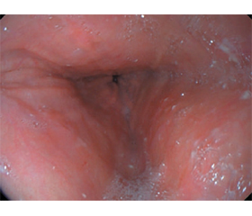
Актуальність. Останнім часом для оцінки фізіологічної кардії при ахалазії стравоходу (АС) використовується метод ендоскопічної ультрасонографії (ЕУС), що поєднує можливості двох досліджень (ендоскопічного та ультразвукового) додатково до традиційних методів діагностики (рентгенографія та ендоскопія) та дає можливість виявити структурні зміни стінки стравоходу і нижнього стравохідного сфінктера (НСС). Мета. Визначити роль ЕУС в оцінці стану фізіологічної кардії при ахалазії стравоходу. Матеріали та методи. У відділі хірургії органів травлення Державної установи «Інститут гастроентерології НАМН України» в 2023–2024 рр., згідно із завданнями роботи, проведено дослідження у 15 пацієнтів з АС та у 20 практично здорових осіб (контрольна група). Усім пацієнтам виконували рентгенологічне та ендоскопічне дослідження стравоходу, шлунка, дванадцятипалої кишки, ендоскопічну ультрасонографію. Результати. При виконанні відеоезофагогастродуоденоскопії виявлено зміни, що характерні для АС: розширення діаметра стравоходу (100,0 %), застійний вміст у просвіті стравоходу (66,7 %), опір при проходженні ендоскопом НСС (86,7 %). При виконанні рентгенологічного дослідження за перистальтикою і діаметром стравоходу встановлено АС І стадії у 13,3 % випадків, ІІ стадії — у 33,3 % випадків, ІІІ стадії — у 46,7 % випадків, ІV стадії — у 6,7 % випадків. При виконанні ЕУС-сканування стравохідний отвір діафрагми становив 19,29 мм (норма 23–36 мм); товщина стінки стравоходу у середній третині — 6,76 мм (норма < 3 мм); товщина стінки НСС — 6,02 мм (норма < 5 мм). Для виявлення фіброзних змін стінки стравоходу та стінки НСС застосовували режим компресійної еластографії. За допомогою компресійної еластографії у 46,6 % випадків виявлено значне потовщення внутрішнього циркулярного м’яза НСС від 2,5 до 4,5 мм, у 26,6 % випадків при АС ІІІ та ІV стадії виявлено фіброзні зміни нижньої третини стінки стравоходу. Висновки. Встановлено, що ЕУС при АС дає відповіді на питання, на які не відповідають традиційні методи дослідження, а саме дає можливість оцінити низку параметрів (товщину стінок НСС та нижньої третини стравоходу, діаметр стравохідного отвору діафрагми) та виявити структурні зміни (наявність фіброзу), що значно впливає на вибір способу хірургічної корекції фізіологічної кардії при АС.
Background. Recently, to assess the physiological cardia in esophageal achalasia (EA), the method of endoscopic ultrasonography (EUS) has been used, which combines the capabilities of two studies, endoscopic and ultrasound, in addition to traditional diagnostic methods, radiography and endoscopy, and allows detecting structural changes in the esophageal wall and lower esophageal sphincter (LES). Objective: to determine the role of endoscopic ultrasonography in assessing the state of physiological cardia in esophageal achalasia. Materials and methods. In the Department of Digestive Surgery of the SI “Institute of Gastroenterology of the National Academy of Medical Sciences of Ukraine” in 2023–2024, according to the objectives of the study, 15 patients with EA and 20 practically healthy individuals (control group) were examined. All patients underwent X-ray and endoscopic examination of the oesophagus, stomach, duodenum, and EUS. Results. Videoesophagogastroduodenoscopy revealed changes characteristic of EA: dilation of the esophageal diameter (100.0 %), stagnant contents in the esophageal lumen (66.7 %), resistance to the passage of an endoscope through LES (86.7 %). X-ray revealed EA stage I in 13.3 % of cases by peristalsis and esophageal diameter, stage II — in 33.3 %, stage III — in 46.7 %, and stage IV — in 6.7 % of cases. When performing EUS, the esophageal hiatus was 19.29 mm (normal 23–36 mm); the esophageal wall thickness in the middle third was 6.76 mm (normal < 3 mm); the LES wall thickness was 6.02 mm (normal < 5 mm). To detect fibrotic changes in the esophageal and the LES wall, compression elastography was used. This method helped reveal a significant thickening of the internal circular muscle of the LES from 2.5 to 4.5 mm in 46.6 % of cases, and in 26.6 %, with EA stage III and IV, fibrotic changes were detected in the lower third of the esophageal wall. Conclusions. It has been found that EUS in EA answers questions that are not answered by traditional research methods, namely, it makes it possible to assess a number of parameters (thickness of the walls of the LES and the lower third of the esophagus, diameter of the esophageal hiatus) and identify structural changes (the presence of fibrosis), which significantly affects the choice of a method for a surgical correction of physiological cardia in EA.
ахалазія стравоходу, ендоскопічне ультразвукове дослідження; компресійна еластографія
esophageal achalasia; endoscopic ultrasound; compression elastography
Для ознакомления с полным содержанием статьи необходимо оформить подписку на журнал.
- Pesce M., Pagliaro M., Sarnelli G., Sweis R. Modern Achalasia: Diagnosis, Classification, and Treatment. J Neurogastroenterol Motil. 2023. № 29(4). Р. 419-427. doi: 10.5056/jnm23125. PMID: 37814432; PMCID: PMC10577462.
- Achalasia / E. Savarino et al. Nat Rev Dis Primers. 2022. № 8(1). 28 р. doi: 10.1038/s41572-022-00356-8. PMID: 35513420.
- Pomenti S., Blackett J.W., Jodorkovsky D. Achalasia: Diagnosis, Management and Surveillance. Gastroenterol Clin North Am. 2021. № 50(4). Р. 721-736. doi: 10.1016/j.gtc.2021.07.001. Epub 2021 Oct 2. PMID: 34717867.
- Achalasia, from diagnosis to treatment / M. Ribolsi et al. Expert Rev Gastroenterol Hepatol. 2023. № 17(1). Р. 21-30. doi: 10.1080/17474124.2022.2163236. PMID: 36588469.
- Cappell M.S., Stavropoulos S.N., Friedel D. Updated Systema–tic Review of Achalasia, with a Focus on POEM Therapy. Dig Dis Sci. 2020. № 65(1). Р. 38-65. doi: 10.1007/s10620-019-05784-3. PMID: 31451984.
- Kahrilas P.J., Ghosh S.K., Pandolfino J.E. Esophageal motility disorders in terms of pressure topography: the Chicago classification. J Clin. Gastroenterol. 2008. Vol. 42. P. 627-635.
- Blonski W., Kumar A., Feldman J., Richter J.E. Timed barium swallow: diagnostic role and predictive value in untreated achalasia, esophagogastric junction outflow obstruction, and non-achalasia dysphagia. Am J Gastroenterol. 2018. № 113(2). Р. 196-203.
- Esophageal motility disorders on high-resolution manometry: Chicago classification version 4.0© / R. Yadlapati et al. Neurogastroenterol Motil. 2021. № 33(1). e14058.
- Present status and perspectives of endosonography 2017 in gastroenterology / M. Hocke et al. Korean J Intern Med. 2018. № 33(1). P. 36-63. doi: 10.3904/kjim.2017.212.
- Iglesias-Garcia J., de la Iglesia-Garcia D., Lariño-Noia J., Dominguez-Muñoz J.E. Endoscopic Ultrasound (EUS) Guided Elastography. Diagnostics. 2023. № 13 (10). P. 1686. https://doi.org/10.3390/diagnostics13101686.
- Ендоскопічна ультразвукова сонографія в діагностиці патології шлунково-кишкового тракту / Ю.М. Степанов та ін. Гастроентерологія. 2021. № 3(55). С. 62-68. DOI: https://doi.org/10.22141/2308-2097.55.3.2021.241590.
- Do we need elastography for EUS? / C.F. Dietrich et al. Endosc Ultrasound. 2020. № 9(5). Р. 284-290. doi: 10.4103/eus.eus_25_20. PMID: 32675464; PMCID: PMC7811716.
- Endoscopic ultrasonography: Enhancing diagnostic accuracy / J. Iglesias-Garcia et al. Best Pract Res Clin Gastroenterol. 2022. № 60-61. Р. 101808. doi: 10.1016/j.bpg.2022.101808.
- Clinical and financial outcomes of per-oral endoscopic myo–tomy compared to laparoscopic heller myotomy for treatment of achalasia / L. Shally et al. Surg Endosc 2022. https://doi.org/10.1007/s00464-022-09652-6.
- Use and Safety of Per-Oral Endoscopic Myotomy for Achalasia in the US / A.W. Lois et al. JAMA Surg. 2022 № 157(6). Р. 490-497. doi: 10.1001/jamasurg.2022.0807. PMID: 35442413; PMCID: PMC9021980.
- Comparison of costs and short-term clinical outcomes of per-oral endoscopic myotomy and laparoscopic Heller myotomy / A. Wirsching et al. Am J Surg. 2019. № 218(4) Р. 706-711. doi: 10.1016/j.amjsurg.2019.07.026. PMID: 31353034.
- Ендоскопія травного тракту. Норма, патологія, сучасні класифікації / В.Й. Кімакович та ін. Львів: Медицина Світу, 2008. 208 с. ISBN 978-966-7475-22-2.
- ASGE guideline on the management of achalasia / M.A. Khashab et al. Gastrointest Endosc. 2020. № 91(2). Р. 213-227.e6. doi: 10.1016/j.gie.2019.04.231. PMID: 31839408.
- Comparing cost and outcomes between peroral endoscopic myo–tomy and laparoscopic heller myotomy / M. Attaar et al. Am J Surg. 2021 № 222(1). Р. 208-213. doi: 10.1016/j.amjsurg.2020.10.037. PMID: 33162014.
- Systematic Review and Bayesian Network Meta-Analysis Comparing Laparoscopic Heller Myotomy, Pneumatic Dilatation, and Peroral Endoscopic Myotomy for Esophageal Achalasia / A. Aiolfi et al. J Laparoendosc Adv Surg Tech A. 2020. № 30(2). Р. 147-155. doi: 10.1089/lap.2019.0432. PMID: 31364910.
- Dirks R.C., Kohn G.P., Slater B. SAGES guidelines committee. Is peroral endoscopic myotomy (POEM) more effective than pneumatic dilation and Heller myotomy? A systematic review and meta-analysis. Surg Endosc. 2021. № 35(5). Р. 1949-1962. doi: 10.1007/s00464-021-08353-w. PMID: 33655443.
- Endoscopic ultrasound-measured muscular thickness of the lo–wer esophageal sphincter and long-term prognosis after peroral endoscopic myotomy for achalasia / E. Liao et al. World J Gastroenrerology. 2020. Oct. 14. № 26(38). Р. 5863-5873. doi: 10.3748/wjg. V26.i38.5863.