Международный эндокринологический журнал Том 20, №7, 2024
Вернуться к номеру
Зміни поляризації M1/M2-макрофагів печінки при довготривалому введенні триптореліну в щурів
Авторы: M.V. Rud, Y.V. Stetsuk, V.I. Shepitko, O.V. Vilkhova, L.B. Pelypenko, O.V. Voloshyna, A.G. Sydorenko, H.P. Pavlenko, N.M. Sharlai, O.V. Sych
Poltava State Medical University, Poltava, Ukraine
Рубрики: Эндокринология
Разделы: Клинические исследования
Версия для печати
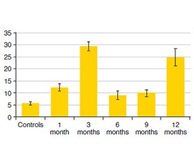
Актуальність. Макрофаги печінки відіграють ключову роль у підтримці гомеостазу цього органа й всього організму, їх можна розділити на три категорії залежно від походження. Макрофаги М1, що позначаються як класично активовані макрофаги, відрізняються від макрофагів, стимульованих інтерфероном гамма або лігандами Toll-подібних рецепторів. Клітини М2 походять від макрофагів, що були стимульовані IL-4/13. Рак передміхурової залози є другою за поширеністю злоякісною пухлиною серед чоловіків у світі. Андроген-деприваційна терапія із використанням агоніста гонадотропін-рилізинг-гормону залишається основою сучасного лікування раку передміхурової залози. Мета: з’ясувати вплив пригнічення тестостерону на імунокомпетентні клітини печінки в самців щурів. У роботі застосовувалася серія експериментальних періодів із введенням триптореліну й кверцетину на різних стадіях. Матеріали та методи. Дослідження проведено на 35 статевозрілих щурах-самцях. Випадковим чином їх розподілили на дві групи: контрольну (n = 10) та експериментальну (n = 25). Тваринам експериментальної групи вводили розчин триптореліну в дозі 0,3 мг активної речовини на 1 кг маси тіла з метою модуляції центральної депривації синтезу лютеїнізуючого гормону. Використано первинні антитіла проти CD163 та CD68. Результати. У печінці тварин на 30-ту добу експерименту кількість клітин CD68+ становила 12,2400 ± 1,5792 у полі зору (p < 0,01), що в 2,2 раза більше, ніж у контрольній групі. Експресія CD163+ зі значенням 25,04 ± 1,79 (p < 0,01) була в 5,56 раза вище порівняно з контрольною групою. На 90-ту добу кількість клітин з експресією CD68+ та CD163+ становила відповідно 29,34 ± 1,86 та 25,66 ± 4,22 (p < 0,01), що в 5,46 разa більше показника в контрольній групі. На 180-й день спостереження визначалося зниження експресії CD68+ порівняно з попереднім періодом обстеження, а експресія CD163+ в клітинах, навпаки, була підвищеною на 40 %. На 270-ту добу експерименту продемонстровано поступове збільшення експресії клітин із CD68+ (9,86 ± 1,47; p < 0,05). Висновки. Тривале застосування триптореліну викликає кількісні та якісні модифікації популяції макрофагів. Максимальна кількість клітин із фенотипом М1 виявляється на 3-му й 12-му місяці спостереження. Кількість фенотипу М2 збільшується на 6-му місяці з поступовим зменшенням до 12-го місяця.
Background. Liver macrophages play a pivotal role in maintaining the homeostasis of the liver and the entire body, they may be classified into three categories based on their origin. M1 macrophages, designated as classically activated macrophages, differentiate from macrophages stimulated by interferon-gamma or ligands of Toll-like receptors. M2 cells are derived from macrophages that have been stimulated by IL-4/13. Prostate cancer is the second most prevalent malignancy in men globally. Androgen deprivation therapy using gonadotropin-releasing hormone agonist remains the backbone of advanced prostate cancer treatment. The purpose of our study was to ascertain the impact of testosterone suppression on immunocompetent hepatic cells in male rats. The study employed a series of experimental periods, with the introduction of triptorelin and quercetin at varying stages. Materials and methods. The study was conducted on 35 sexually mature male rats. They were randomly allocated to two groups: the controls (n = 10) and the experimental one (n = 25). The animals in the experimental group were administered a solution of triptorelin at a dose of 0.3 mg of active ingredient per 1 kg of body weight, with the aim of modulating the central deprivation of luteinizing hormone synthesis. We used primary antibodies against CD163 and CD68. Results. In the livers of animals on day 30 of the experiment, the number of CD68+ cells were calculated to be 12.2400 ± 1.5792 per FOV at p < 0.01, which is 2.2 times higher than in the control group. The expression of CD163+ with a value of 25.04 ± 1.79 (p < 0.01) is 5.56 times higher compared to the controls. On day 90, the number of cells exhibiting CD68+ and CD163+ expression was 29.34 ± 1.86 and 25.66 ± 4.22, respectively, at p < 0.01, representing a 5.46-fold increase compared to the FOV in the control group preparations. The 180th day of observation was defined by a reduction in the expression of CD68+ as compared to the earlier examination period. Conversely, the expression of CD163+ in cells increased, representing a 40% rise compared to the previous examination period. On day 270 of the experiment, a gradual increase in the expression of cells with CD68+ (9.86 ± 1.47; p < 0.05) was demonstrated. Conclusions. The long-term administration of triptorelin causes changes in quantitative and qualitative modifications of the macrophage population. Maximum of M1 phenotype are detected at 3 and 12 months of observation. The number of M2 phenotype increases by 6 months of monitoring, with a gradual decrease by 12 months.
печінка; макрофаги; трипторелін; CD68; CD163; лютеїнізуючий гормон
liver; macrophages; triptorelin; CD68; CD163; luteinizing hormone
Для ознакомления с полным содержанием статьи необходимо оформить подписку на журнал.
- Krenkel O, Tacke F. Liver macrophages in tissue homeostasis and disease. Nat Rev Immunol. 2017 May;17(5):306-321. doi: 10.1038/nri.2017.11.
- Ahamed F, Eppler N, Jones E, Zhang Y. Understanding Macrophage Complexity in Metabolic Dysfunction-Associated Steatotic Liver Disease: Transitioning from the M1/M2 Paradigm to Spatial Dynamics. Livers. 2024 Sep;4(3):455-478. doi: 10.3390/livers4030033.
- Gordon S. Alternative activation of macrophages. Nat Rev Immunol. 2003 Jan;3(1):23-35. doi: 10.1038/nri978.
- Mosser DM, Edwards JP. Exploring the full spectrum of macrophage activation. Nat Rev Immunol. 2008 Dec;8(12):958-69. doi: 10.1038/nri2448.
- Cai Z, Xie Q, Hu T, Yao Q, Zhao J, Wu Q, Tang Q. S100A8/A9 in Myocardial Infarction: A Promising Biomarker and Therapeutic Target. Front Cell Dev Biol. 2020 Nov 12;8:603902. doi: 10.3389/fcell.2020.603902.
- Sica A, Mantovani A. Macrophage plasticity and polarization: in vivo veritas. J Clin Invest. 2012 Mar;122(3):787-95. doi: 10.1172/JCI59643.
- Muraille E, Leo O, Moser M. 2014. Th1/Th2 paradigm extended: macrophage polarization as an unappreciated pathogen-dri–ven escape mechanism? Front Immunol. 2014;5:603. doi: 10.3389/fimmu.2014.00603.
- Bai L, Kong M, Duan Z, et al. M2-like macrophages exert hepatoprotection in acute-on-chronic liver failure through inhibi–ting necroptosis-S100A9-necroinflammation axis. Cell Death Dis. 2021;12:93. doi: 10.1038/s41419-020-03378-w.
- Zhou H, Zhao C, Shao R, Xu Y, Zhao W. The functions and re–gulatory pathways of S100A8/A9 and its receptors in cancers. Front Pharmacol. 2023 Aug 28;14:1187741. doi: 10.3389/fphar.2023.1187741.
- Kerkhoff C, Klempt M, Sorg C. Novel insights into structure and function of MRP8 (S100A8) and MRP14 (S100A9). Biochim Biophys Acta. 1998 Dec 10;1448(2):200-11. doi: 10.1016/s0167-4889(98)00144-x.
- Akimov OY, Kostenko VO. Functioning of nitric oxide cycle in gastric mucosa of rats under excessive combined intake of sodium nitrate and fluoride. Ukr Biochem J. 2016 Nov-Dec;88(6):70-5. doi: 10.15407/ubj88.06.070.
- Raja T, Sud R, Addla S, Sarkar KK, Sridhar PS, et al. Gonadotropin-releasing hormone agonists in prostate cancer: A comparative review of efficacy and safety. Indian J Cancer. 2022 Mar;59 (Suppl):S142-S159. doi: 10.4103/ijc.IJC_65_21.
- Montagnani Marelli M, Moretti RM, Januszkiewicz-Cau–lier J, Motta M, Limonta P. Gonadotropin-releasing hormone (GnRH) receptors in tumors: a new rationale for the therapeutical application of GnRH analogs in cancer patients? Curr Cancer Drug Targets. 2006 May;6(3):257-69. doi: 10.2174/156800906776842966.
- Spitz A, Young JM, Larsen L, Mattia-Goldberg C, Donnelly J, Chwalisz K. Efficacy and safety of leuprolide acetate 6-month depot for suppression of testosterone in patients with prostate cancer. Prostate Cancer Prostatic Dis. 2012 Mar;15(1):93-9. doi: 10.1038/pcan.2011.50.
- Kur P, Kolasa-Wołosiuk A, Misiakiewicz-Has K, Wiszniewska B. Sex Hormone-Dependent Physiology and Diseases of Liver. Int J Environ Res Public Health. 2020 Apr 11;17(8):2620. doi: 10.3390/ijerph17082620.
- Song MJ, Choi JY. Androgen dysfunction in non-alcoholic fatty liver disease: Role of sex hormone binding globulin. Front Endocrinol (Lausanne). 2022 Nov 22;13:1053709. doi: 10.3389/fendo.2022.1053709.
- Lin HY, Yu IC, Wang RS, Chen YT, Liu NC, et al. Increased hepatic steatosis and insulin resistance in mice lacking hepatic androgen receptor. Hepatology. 2008;47:1924-1935. doi: 10.1002/hep.22252.
- Chen R, Kang R, Tang D. The mechanism of HMGB1 secretion and release. Exp Mol Med. 2022 Feb;54(2):91-102. doi: 10.1038/s12276-022-00736-w.
- Yang H, Wang H, Andersson U. Targeting Inflammation Dri–ven by HMGB1. Front Immunol. 2020 Mar 20;11:484. doi: 10.3389/fimmu.2020.00484.
- Fang P, Liang J, Jiang X, Fang X, Wu M, et al. Quercetin Attenuates d-GaLN-Induced L02 Cell Damage by Suppressing Oxidative Stress and Mitochondrial Apoptosis via Inhibition of HMGB1. Front Pharmacol. 2020 May 5;11:608. doi: 10.3389/fphar.2020.00608.
- Chekalina NI, Kazakov YM, Mamontova TV, Vesnina LE, Kaidashev IP. Resveratrol more effectively than quercetin reduces endothelium degeneration and level of necrosis factor α in patients with coronary artery disease. Wiad Lek. 2016;69(3, pt 2):475-479.
- Stetsuk YeV, Shepitko VI, Akimov OYe, Yakushko OS, Solo–vyova NV. Influence of quercetin on morphological changes in rats testes after 180 days during central deprivation of luteinizing hormone. World of Medicine and Biology. 2021;3(77):243-248. doi: 10.26724/2079-8334-2021-3-77-243-248.
- Martynenko R, Shepitko V, Stetsuk Y, Boruta N, Rud M, et al. Expression of Ki67 and CD68+ cells of red bone marrow monocyte sprout under triptorelin administration in the hypothalamic-pituitary-testis regulatory system: the experimental study. Internatio–nal Journal of Endocrinology (Ukraine). 2023;19(6):412-418. doi: 10.22141/2224-0721.19.6.2023.1308.
- Stetsuk YV, Shepitko VI, Pronina OM, Zaporozhets TM, Boruta NV, et al. Effect of quercetin administration on electron microscopic changes in testicular interstitial endocrinocytes during long-term central blockade of luteinising hormone in rats. Reports of Morphology. 2024;30(1):68-75. doi: 10.31393/morphology-journal-2024-30(1)-09.
- Kolomiiets O, Moskalenko R. Immunohistochemical study of m1 and m2 macrophages in breast cancer with microcalcifications. East Ukr Med J. 2023;11(2):155-63. doi: 10.21272/eumj.2023;11(2):155-163.
- Cheng D, Chai J, Wang H, Fu L, Peng S, Ni X. Hepatic macrophages: Key players in the development and progression of liver fibrosis. Liver Int. 2021 Oct;41(10):2279-2294. doi: 10.1111/liv.14940.
- Plevriti A, Lamprou M, Mourkogianni E, Skoulas N, Giannakopoulou M, et al. The Role of Soluble CD163 (sCD163) in Human Physiology and Pathophysiology. Cells. 2024 Oct 11;13(20):1679. doi: 10.3390/cells13201679.
- Nielsen MC, Hvidbjerg Gantzel R, Clària J, Trebicka J, Møller HJ, Grønbæk H. Macrophage Activation Markers, CD163 and CD206, in Acute-on-Chronic Liver Failure. Cells. 2020 May 9;9(5):1175. doi: 10.3390/cells9051175.
- Rahman N, Pervin M, Kuramochi M, Karim MR, Izawa T, Kuwamura M, Yamate J. M1/M2-macrophage Polarization-based Hepatotoxicity in d-galactosamine-induced Acute Li–ver Injury in Rats. Toxicol Pathol. 2018 Oct;46(7):764-776. doi: 10.1177/0192623318801574.
- Grønbæk H, Rødgaard-Hansen S, Aagaard NK, Arroyo V, Moestrup SK, et al.; CANONIC study investigators of the EASL-CLIF Consortium. Macrophage activation markers predict mortality in patients with liver cirrhosis without or with acute-on-chronic liver failure (ACLF). J Hepatol. 2016 Apr;64(4):813-22. doi: 10.1016/j.jhep.2015.11.021.

