Журнал «Здоровье ребенка» Том 19, №8, 2024
Вернуться к номеру
Сторонні тіла верхніх відділів травного каналу в дітей, сучасні підходи до видалення. Клінічний випадок
Авторы: Іванців В.А. (1, 2), Няньковський С.Л. (2), Ющик Л.В. (2), Няньковська О.С. (2), Тумак І.М. (3), Кочеркевич Т.О. (1), Юрків О.В. (1), Кочеркевич О.Н. (4)
(1) - ВП «Лікарня Святого Миколая», Перше територіальне медичне об’єднання міста Львова, м. Львів, Україна
(2) - Львівський національний медичний університет імені Данила Галицького, м. Львів, Україна
(3) - ВП «Лікарня Святого Пантелеймона», Перше територіальне медичне об’єднання міста Львова,
м. Львів, Україна
(4) - Західноукраїнський спеціалізований дитячий медичний центр, м. Львів, Україна
Рубрики: Педиатрия/Неонатология
Разделы: Справочник специалиста
Версия для печати
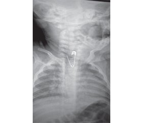
Актуальність. Проблема надання невідкладної допомоги дітям, які ковтають гострі сторонні предмети, залишається актуальною і знаходиться в площині вирішення таких питань: у які терміни надається така допомога, який пошуковий алгоритм визначення характеру стороннього тіла (СТ) і які методичні аспекти техніки проведення самої маніпуляції лікарем-ендоскопістом. Мета: удосконалення методики видалення фіксованого гостро-колючого предмета зі стравоходу в дітей. Матеріали та методи. У статті описаний клінічний випадок ендоскопічного видалення розкритої шпильки довжиною 2 см гострим кінцем догори з вклиненням у першому фізіологічному звуженні стравоходу. Результати. Ендоскопічна операція видалення гостро-колючого стороннього тіла з вклиненням в стравоході проводилась в три етапи. Перший етап складався з послідовного та безпечного опускання розкритої шпильки в шлунок для її переорієнтації гострим кінцем донизу для уникнення пошкодження стінок кардії стравоходу та його гирла на виході. Другий етап полягав у розвертанні та переорієнтації голівки шпильки гострим кінцем донизу. Третій етап — безпечна й надійна фіксація СТ гострим кінцем донизу та його повільне виведення назовні. Висновки. Обстеження й лікування хворих дітей з підозрою на СТ верхніх відділів травного каналу повинно здійснюватися в екстреному порядку в умовах хірургічного відділення спеціалізованого багатопрофільного стаціонару. Оптимальною є централізація такої допомоги в одній установі області з відповідним забезпеченням її спеціалізованим обладнанням та інструментарієм. Будь-яке виявлене в просвіті верхніх відділів травного каналу СТ повинно бути видаленим, по змозі, за допомогою гнучкої ендоскопії. Для діагностики характеру СТ і вивчення причини фіксації його в просвіті необхідне поєднання рентгенологічного й ендоскопічного методів дослідження. З метою мінімізації ризику перфорації стінки порожнистого органа ендоскопічне втручання в дітей слід обов’язково проводити під загальним знечуленням в умовах операційної. Застосування гнучких ендоскопів відповідного типу з урахуванням вікових анатомо-фізіологічних особливостей мінімізує травматизацію стінок стравоходу і зводить до мінімуму ризик перфорації стінки порожнистого органа.
Background. The problem of providing emergency care to children who have swallowed sharp foreign bodies remains relevant and requires addressing the following issues: the timeliness of such care, the diagnostic algorithm for determining the nature of a foreign body, and the methodological aspects of the endoscopist’s procedure. The purpose of this study is to improve the technique for removing a fixed sharp-pointed foreign body from a child’s esophagus. Materials and methods. The article describes a clinical case of endoscopic removal of a 2-cm long open safety pin lodged in the upper esophageal sphincter with the sharp end up. Results. The endoscopic procedure to remove an impacted sharp foreign body from the esophagus was conducted in three steps. The first stage involved the sequential and safe moving the open safety pin to the stomach to reorient it with the sharp end pointing downward, thereby preventing injury to the cardiac sphincter and the esophageal orifice during its removal. The second stage involved rotating and reorienting the safety pin so that the sharp end pointed downward. The third stage involved the secure fixation of a foreign body with the sharp end pointing downward and its slow, careful removal. Conclusions. Children suspected of having a foreign body in the upper gastrointestinal tract should be urgently assessed and treated in the surgical department of a specialized multidisciplinary hospital. The most effective way to provide this type of care is to centralize it in a single regional facility that is fully equipped with specialized equipment and instruments. Flexible endoscopy is the preferred method for removal of foreign bodies found in the upper gastrointestinal tract. A combination of radiographic and endoscopic examinations is necessary to determine the nature of a foreign body and to investigate the cause of its fixation in the lumen. To prevent perforation of the hollow organ, endoscopic procedures in children must be carried out under general anesthesia in a surgical setting. The use of appropriate flexible endoscopes, considering the age-related anatomical and physiological characteristics, minimizes trauma to the esophageal wall and reduces the risk of perforation of the hollow organ wall.
сторонні тіла; діти; стравохід; ендоскопія; невідкладна допомога
foreign bodies; children; esophagus; endoscopy; emergency care
Для ознакомления с полным содержанием статьи необходимо оформить подписку на журнал.
- Tringali A, Thomson M, Dumonceau JM, Tavares M, Tabbers MM, Furlano R, et al. Pediatric gastrointestinal endoscopy: European Society of Gastrointestinal Endoscopy (ESGE) and European Society for Paediatric Gastroenterology Hepatology and Nutrition –(ESPGHAN) guideline executive summary. Endoscopy. 2017;49(1):83-91. doi: 10.1097/MPG.0000000000001408.
- Cagil Y, Diaz J, Iskowitz S, Muñiz Crim AJ. Ingested Foreign Bodies and Toxic Materials: Who Needs to be Scoped and When? Pediatrics in Review. 2021 Jun;42(6):290-301. doi: https://doi.org/10.1542/pir.2018-0327.
- Speidel AJ, Wölfle L, Mayer B, et al. Increase in foreign body and harmful substance ingestion and associated complications in children: a retrospective study of 1199 cases from 2005 to 2017. BMC Pediatr. 2020;20:560. [PubMed]. doi: https://doi.org/10.1186/s12887-020-02444-8.
- Novotny EW, Keel PC. Swallowed Foreign Bodies that have Cardiac Complications in Children. Pediatric Care. 2021;7(1):5. doi: 10.36648/2471-805X.7.1.64.
- Yuan J, Ma M, Guo Y, He B, Cai Z, Ye B, et al. Delayed endoscopic removal of sharp foreign body in the esophagus increased clinical complications: an experience from multiple centers in China. Medicine. 2019;98(26):16146. doi: 10.1097/MD.0000000000016146.
- Conners GP. Pediatric Foreign Body Ingestion Clinical Presentation. Updated: Mar 03; 2023. https://emedicine.medscape.com/article/801821-clinical.
- Khorana J, Tantivit Y, Phiuphong C, Pattapong S, Siripan S. Foreign Body Ingestion in Pediatrics: Distribution, Management and Complications. Medicina (Kaunas). 2019 Oct;55(10):686. doi: 10.3390/medicina55100686.
- Lee JH, Lee JH, Shim JO, Lee JH, Eun B-L, Yoo KH. Fo–reign Body Ingestion in Children: Should Button Batteries in the Sto–mach Be Urgently Removed? Pediatr Gastroenterol Hepatol Nutr. 2016 Mar;19(1):20-28. doi: 10.5223/pghn.2016.19.1.20.
- Au А, Goldman RD. Management of gastric metallic foreign bodies in children. Can Fam Physician. 2021 Jul;67(7):503-505. doi: 10.46747/cfp.6707503.
- Lee JH. Foreign Body Ingestion in Children. Clin Endosc. 2018;51(2):129-136. doi: https://doi.org/10.5946/ce.2018.039.
- Orsagh-Yentis D, McAdams RJ, Roberts KJ, McKenzie LB. Foreign-body ingestions of young children treated in US emergency departments: 1995–2015. Pediatrics. 2019;143(5):2018-1988. doi: https://doi.org/10.1542/peds.2018-1988.
- Yeh H-Y, Chao H-Ch, Chen Sh-Y, Chen Ch-Ch, Lai M-W. Analysis of Radiopaque Gastrointestinal Foreign Bodies Expelled by Spo–ntaneous Passage in Children: A 15-Year Single-Center Study. Front Pediatr. 2018 June:1-9. doi: https://doi.org/10.3389/fped.2018.00172.
- Dipasquale V, Romano С, Iannelli М, Tortora A, Melita G, Ventimiglia М, Pallio S. Managing Pediatric Foreign Body Ingestions: A –10-Year Experience. Pediatr Emerg Care. 2022 Jan 1;38(1):e268-e271. doi: 10.1097/PEC.0000000000002245.
- Ma Т, Zheng W, An В, Xia Y, Chen G. Small bowel perforation secondary to foreign body ingestion mimicking acute appendicitis. Case report. Medicine (Baltimore). 2019 Jul;98(30):164-89. [PubMed]. doi: 10.1097/MD.0000000000016489.
- Kroon HM, Mullen D. Ingested foreign body causing a silent perforation of the bowel. BMJ Case Rep. 2021;14:240879. doi: 10.1136/bcr-2020-240879.
- Vo NQ, Nguyen LD, Chau THT, Tran VK, Nguyen TT. Toothpick — a rare cause of bowel perforation: case report and literature review. Radiology Case Reports. 2020;15(10):1799-1802. doi: https://doi.org/10.1016/j.radcr.2020.07.034.
- Ingraham CR, Mannelli L, Robinson JD, Linnau KF. Radio–logy of foreign bodies: how do we image them? Emergency Radiology. 2015;22:425-430.doi: https://doi.org/10.1007/s10140-015-1294-9.
- Tashtush NA, Bataineh ZA, Yusef DH, Al Quran TM, Rou–san LA, Khasawneh R, Aleshawi AJ, Altamimi EM. Ingested sharp foreign body presented as chronic esophageal stricture and inflammatory mediastinal mass for 113 weeks: Case report. Annals of Medicine and Surgery. 2019 Sept;45:91-4. doi: 10.1016/j.amsu.2019.07.028.
- Gatto A, Angelici S, Di Pangrazio C, Nanni L, Buonsenso D, Para–diso FV, Chiaretti A. The Fakir Child: Clinical Observation or Invasive Treatment? Pediatr Rep. 2020;12:103-107. doi: 10.3390/pediatric12030023.
- Hong KH, Kim YJ, Kim JH, Chun SW, Kim HM, Cho JH. Risk factors for complications associated with upper gastrointestinal foreign bodies. World J Gastroenterol. 2015;21(26):8125-8131. doi: 10.3748/wjg.v21.i26.8125.
- Ruan W-Sh, Li Y-N, Feng M-X, Lu Y-Q. Retrospective observational analysis of esophageal foreign bodies: a novel characterization based on shape. Scientific Reports. 2020;10:4273. doi: 10.1038/s41598-020-61207-8.
- Emeka CK, Chukwuebuka NO, Tochukwu EJ. Foreign Body in the Gastrointestinal Tract in Children: A Tertiary Hospital Experience. African Journal of Paediatric Surgery. 2023;20(3):224-228. doi: 10.4103/ajps.AJPS_148_20.
- Green SS. Ingested and Aspirated Foreign Bodies. Pediatrics in Review. 2015;36(10);430-437; doi: https://doi.org/10.1542/pir.36-10-430.
- Qiu Y, Xu Sh, Wang Y, Chen E. Migration of ingested sharp fo–reign body into the bronchus: a case report and review of the literature. BMC Pulm Med. 2021;21:90. https://doi.org/10.1186/s12890-021-01458-x.
- Li С, Yong СС, Encarnacion DD. Duodenal perforation nine months after accidental foreign body ingestion, a case report. BMC Surgery. 2019;19:132. doi: https://doi.org/10.1186/s12893-019-0594-5.
- Zifeng Y, Deqing W, Dailan X, Yong L. Gastrointestinal perforation secondary to accidental ingestion of toothpicks: A series case report. Medicine (Baltimore). 2017 Dec;96(50):e9066. doi: 10.1097/MD.0000000000009066.

